Wide-view-field chromatography hyperspectral microscopic imaging method and device based on space-time focusing

A microscopic imaging and hyperspectral technology, applied in the field of microscopic spectral imaging and analytical chemistry, can solve the problems of spectral imaging of uncomfortable scattering tissue, signal crosstalk, reduction of axial resolution, etc., and achieve low-speed spectral information acquisition, low scattering Effects of signal crosstalk, high temporal resolution

Active Publication Date: 2018-11-06
TSINGHUA UNIV
View PDF5 Cites 19 Cited by
  • Summary
  • Abstract
  • Description
  • Claims
  • Application Information

AI Technical Summary

Problems solved by technology

On the other hand, although nonlinear optical microscopy using surface excitation avoids the speed bottleneck caused by point-by-point scanning, the excited signal has serious crosstalk after being scattered by tissue, which is not suitable for scattering tissue spectral imaging.
Ordinary nonlinear optical microscopy using line-scanning method can compromise between imaging speed and suppression of scattering effects, but this method is not an ideal choice because of the lower axial resolution obtained by point-scanning method

Method used


Image

  • Wide-view-field chromatography hyperspectral microscopic imaging method and device based on space-time focusing
  • Wide-view-field chromatography hyperspectral microscopic imaging method and device based on space-time focusing
  • Wide-view-field chromatography hyperspectral microscopic imaging method and device based on space-time focusing

Examples


Embodiment 1

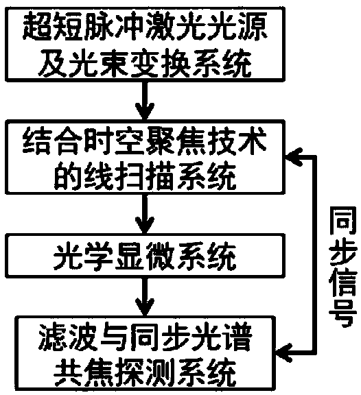
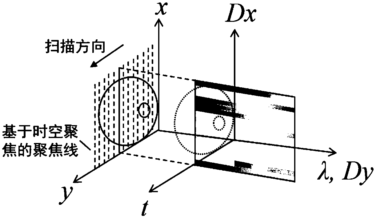
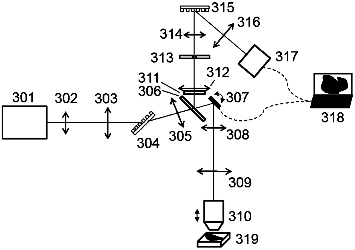
[0049] Refer below image 3 , describe in detail the wide field of view tomographic hyperspectral microscopic imaging device of this embodiment, the device includes an ultrashort pulse laser source and a beam conversion system, a line scanning system based on spatiotemporal focusing, an optical microscopic system, filtering and synchronous spectral sharing focus detection system, the biological sample is placed on the sample stage 319. Wherein, the ultrashort pulse laser source and the ultrashort pulse laser source 301 in the beam conversion system adopt femtosecond lasers (such as Coherent Chameleon Discovery series), and the beam conversion system adopts a Kepler telescope system composed of a lens 302 and a cylindrical lens 303 ( is 4f system); line scanning system based on spatio-temporal focusing includes transmission grating 304, lens 305 and scanning galvanometer 307; optical microscope system includes two lenses 308, 309 and microscope objective lens 310; filtering and...

Embodiment 2

[0052] Refer below Figure 4 , the wide-field tomographic hyperspectral microscopic imaging device of this embodiment is described in detail. The difference between this embodiment and Embodiment 1 is that an adaptive optical element is added. The device in this embodiment includes an ultrashort pulse laser light source and a beam conversion system, a line scanning system based on spatiotemporal focusing, an optical microscope system, a filtering and synchronous spectral confocal detection system, and biological samples are placed on the sample stage 424; wherein, the ultrashort The ultrashort pulse laser source 401 in the pulsed laser light source and the beam conversion system uses a femtosecond laser (such as Coherent Chameleon Discovery series), and the beam conversion system uses a Kepler telescope system composed of a lens 402 and a cylindrical lens 403 (a 4f system) The line scanning system based on spatio-temporal focusing includes a transmission grating 404, five lens...


PUM


Abstract

The invention discloses a wide-view-field chromatography hyperspectral microscopic imaging method and device based on space-time focusing, and belongs to the technical field of microscopic spectral imaging and analytical chemistry. According to the method, ultra-short pulse lasers are generated through an ultra-short pulse laser source, focusing lines are generated in a sample through space-time focusing, excited fluorescence is collected, stray light is filtered out through con-focal optical slits, spectral information (x, lambda), of the sample is acquired through fluorescence spectral information collecting, and finally sample (x, lambda, y, z, t) five-dimensional information is acquired through three-dimensional space scanning and delayed scanning. The device comprises the ultra-shortpulse laser source, a light beam conversion system, a line scanning system based on space-time focusing, an optical microscopic system and a filtering and synchronous spectrum con-focal detection system. Acquisition of the spectral information in the filtering and synchronous spectrum con-focal detection system is synchronous with line scanning trigger signals in the line scanning system in combination with the space-time focusing technology. The method and device have the advantages of being wide in view field, high in space resolution, high in time resolution, high in spectral resolution andthe like.

Description

technical field [0001] The invention relates to a wide field of view tomographic hyperspectral microscopic imaging method and device based on spatiotemporal focusing, and belongs to the technical fields of microspectral imaging and analytical chemistry. Background technique [0002] Hyperspectral Microscopy (Hyperspectral Microscopy) has important applications in the field of biomedical research, especially in the fields of clinical disease diagnosis, intraoperative image navigation and other fields, which have attracted more and more attention. Using hyperspectral microscopic imaging technology to obtain spatially resolved spectral information can provide information on physiological parameters, morphology and components of biological tissues for disease diagnosis. At present, non-invasive detection of various cancers can be realized by using hyperspectral microscopic imaging technology. [0003] In essence, hyperspectral microscopic imaging technology is a technology to o...

Claims


Application Information

Patent Timeline
Patent Type & Authority Applications(China)
IPC IPC(8): A61B5/00
CPCA61B5/0073
Inventor 孔令杰谢浩张元龙戴琼海
Owner TSINGHUA UNIV
Why Eureka
Try Eureka