
Magnetic resonance electromyographic signal trigger

An electromyographic signal and trigger technology, which is used in sensors, medical science, diagnostic recording/measurement, etc., can solve problems such as easy falling off, false triggering, signal instability, etc., to ensure accuracy, small time error, and improve triggering. The effect of precision

Inactive Publication Date: 2012-07-18
THE THIRD AFFILIATED HOSPITAL OF SUN YAT SEN UNIV
View PDF0 Cites 0 Cited by
  • Summary
  • Abstract
  • Description
  • Claims
  • Application Information

AI Technical Summary

Problems solved by technology

[0004] Aiming at the shortcomings of existing myoelectric triggers that convert the trajectory of fluorescent sticks to output digital and analog ECG signals through desktop computers and analog-to-digital conversion software, but the converted signals are unstable, easy to fall off or cause false triggers, the purpose of the present invention is to Provides a small size, easy to install magnetic resonance electromyographic signal trigger, which is more sensitive to the capture of limb movements, generates stable and consistent trigger signals, and ensures the smooth progress of magnetic resonance scanning

Method used


Image

  • Magnetic resonance electromyographic signal trigger
  • Magnetic resonance electromyographic signal trigger
  • Magnetic resonance electromyographic signal trigger

Examples


Embodiment Construction

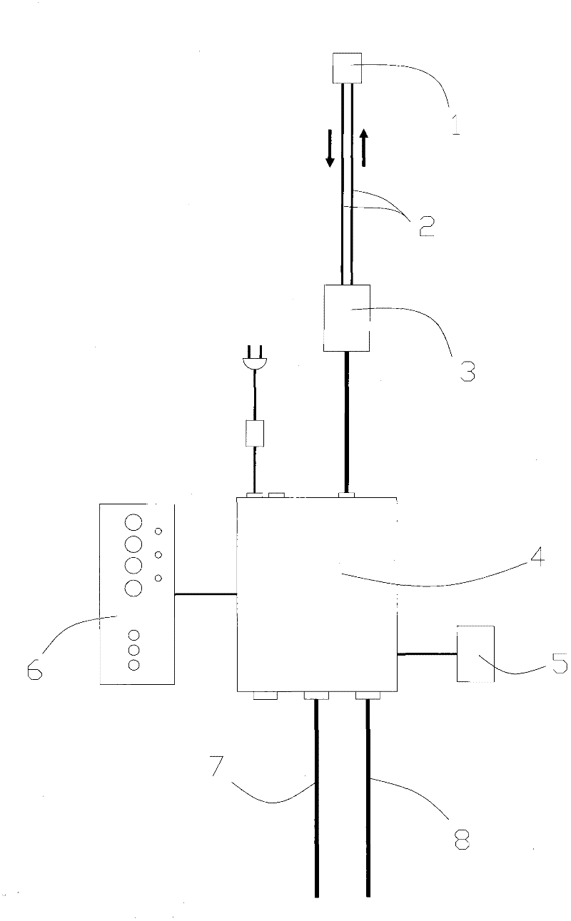
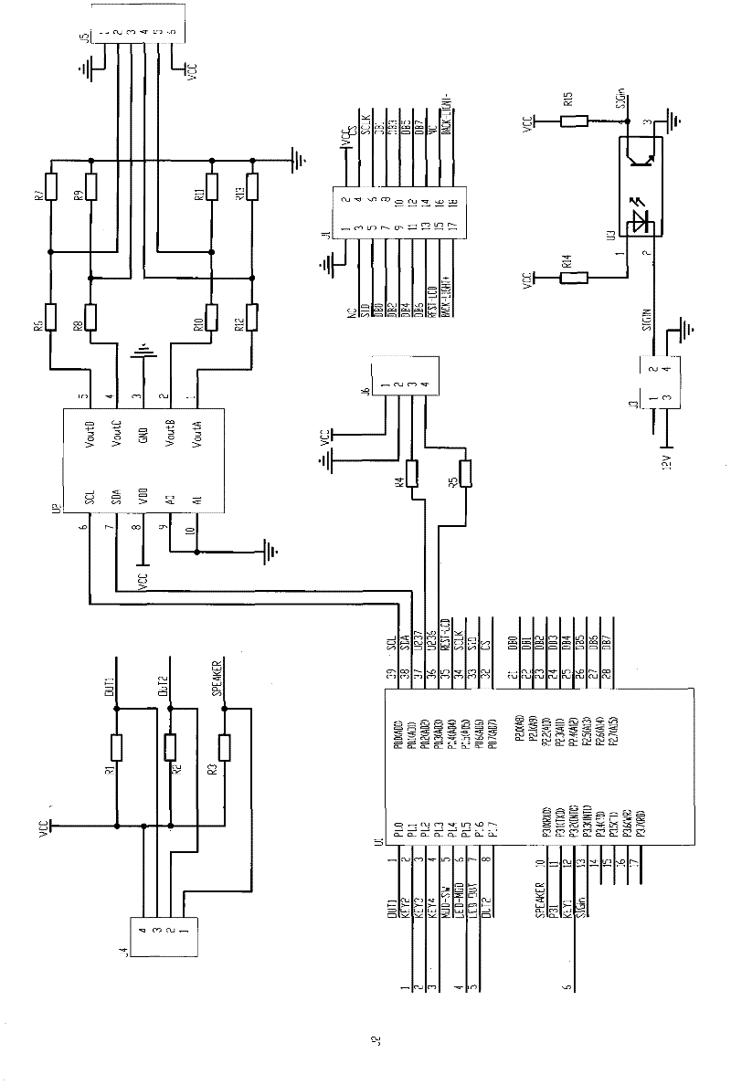
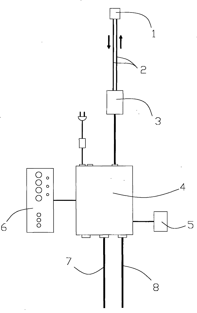
[0016] see figure 1 and figure 2 , the present invention includes main control unit 4, surface electromyography output channel 7, nuclear magnetic resonance instrument output channel 8, liquid crystal display 5 (OAMJ24C), keyboard 6, photoelectric converter 3 (SUNX FX-A1), optical fiber 2 and focusing mirror 1. The photoelectric converter 3 (SUNX FX-A1), the optical fiber 2 and the focusing mirror 1 form a sensor device, the focusing mirror 1 is connected to the optical fiber 2, the optical fiber 2 is connected to the photoelectric converter 3, and the photoelectric converter 3 is connected to the main control unit 4 The output channel 7 of the surface electromyography instrument and the output channel 8 of the nuclear magnetic resonance instrument are connected by the main control unit through the shielded wire, and the output terminals are respectively provided with red copper terminals; the main control unit 4 is connected by the microcontroller U1 (STC89C52), D / A conversi...


PUM


Abstract

The invention discloses a magnetic resonance electromyographic signal trigger, comprising a sensor device, a main control unit, a surface electromyography output channel and a nuclear magnetic resonance spectrometer output channel, wherein the sensor device is connected with the main control unit; and the surface electromyography output channel and the nuclear magnetic resonance spectrometer output channel are respectively connected with the main control unit. The trigger only generates a signal the amplitude of which is greater than the threshold upper limit of Vmax; complicated electrocardio signals can be replaced by sine waves, square, waves or triangular waves and the like; meanwhile, simpler trigger signals can ensure the accuracy of the trigger edge, improve the trigger precision and has smaller time error.

Description

technical field [0001] The invention relates to an image acquisition device, in particular to a magnetic resonance myoelectric signal trigger. Background technique [0002] Velocity-encoded phase-contrast magnetic resonance imaging (VEPC MRI) is a new type of imaging technique for evaluating muscle function in vivo. This technique can be used to acquire functional magnetic resonance imaging of skeletal muscles in moving limbs. image. The trigger is a key device for VEPC MR image acquisition, and the trigger generates a signal to trigger the magnetic resonance scan, thereby completing the image acquisition process. In velocity-encoded phase-contrast MRI technology, EMG triggers are required to collect human movement information in the working environment of the MRI instrument (usually a strong magnetic environment). [0003] At present, there is no such myoelectric trigger product in China. Similar foreign products, such as the Optitrig (model: 2D) of the Finnish NOMIR comp...

Claims


Application Information

Patent Timeline
Patent Type & Authority Patents(China)
IPC IPC(8): A61B5/055A61B5/0488
Inventor 窦祖林白鹏飞李宗涛温红梅姜丽
Owner THE THIRD AFFILIATED HOSPITAL OF SUN YAT SEN UNIV
Learn More