Ultrasound microbubble agent for treating tumors by low-intensity focused ultrasound and preparation method thereof

A technology of focused ultrasound and ultrasonic microbubbles, applied in the field of medicine

Inactive Publication Date: 2011-03-30
INST OF MATERIA MEDICA CHINESE ACAD OF MEDICAL SCI
View PDF0 Cites 4 Cited by
  • Summary
  • Abstract
  • Description
  • Claims
  • Application Information

AI Technical Summary

Problems solved by technology

[0005] However, the particle size of contrast agents currently used clinically is in the range of 2.0-4.5 μm Can only visualize large cardiovascular vessels, but cannot pass through the capillaries in the lesion tissue (the diameter of capillaries in malignant tumors is usually 0.3-1.2μm)

Method used


Image

  • Ultrasound microbubble agent for treating tumors by low-intensity focused ultrasound and preparation method thereof
  • Ultrasound microbubble agent for treating tumors by low-intensity focused ultrasound and preparation method thereof
  • Ultrasound microbubble agent for treating tumors by low-intensity focused ultrasound and preparation method thereof

Examples


Embodiment 1

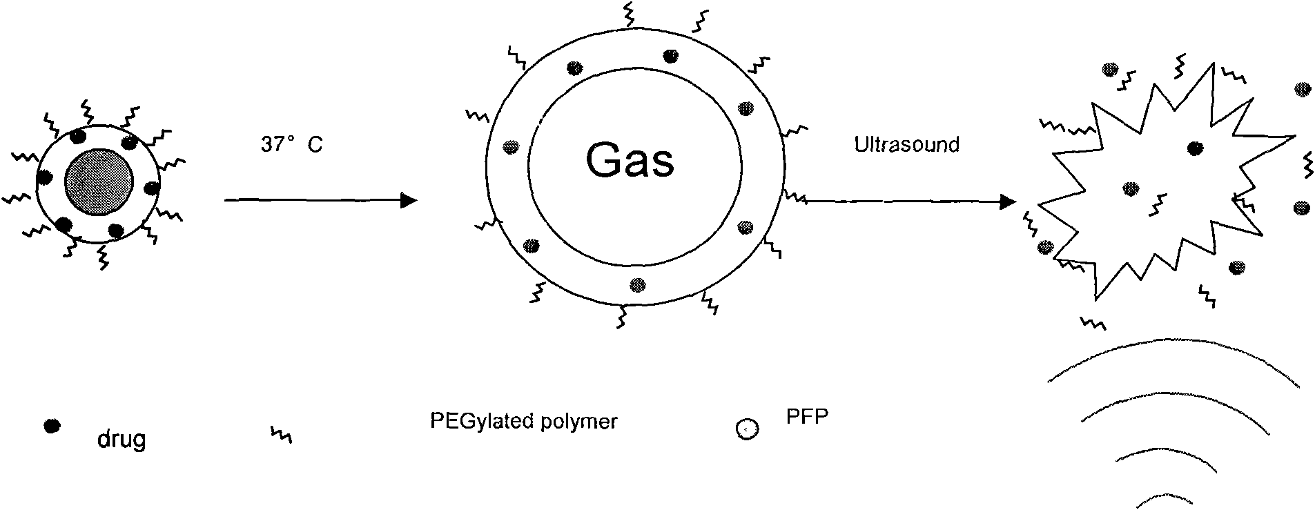
[0028] Nanoscale Ultrasonic Microbubbles Encapsulating Doxorubicin

[0029] The nano-scale ultrasonic microbubble agent wrapped with doxorubicin was prepared according to the following steps:

[0030] In the first step, 50mg PEG 2000 -PCL 2000 and 7.5mg doxorubicin were dissolved in 4ml DMSO, and after complete dissolution, add physiological saline to dilute to 8ml, then put the mixture into a dialysis bag (pore size is 12000Da), dialyze in 100ml physiological saline, change the same amount every day Physiological saline, dialyzed for 2 days, until the organic solvent was completely removed to form a micellar solution containing doxorubicin, followed by filter sterilization (200nm);

[0031] In the second step, 200 mg of filter-sterilized dodecafluoropentane (PFP) was added to the above micellar solution and vortexed for 1 min to obtain a mixed solution;

[0032] The third step is to put the above mixed solution into a sterilized test tube, then place the test tube in an ic...

Embodiment 2

[0034] Nanoscale Ultrasonic Microbubbles Encapsulating Paclitaxel

[0035] Prepare nano-scale ultrasonic microbubbles encapsulating paclitaxel according to the following steps:

[0036] In the first step, 100mg PEG 2000 -PLLA 2000 Dissolve 15mg paclitaxel in 6ml DMF, add water for injection after dissolving completely and dilute to 10ml, then put the mixture into a dialysis bag (pore size is 11000Da), dialyze in 150ml water for injection, change the same amount of water for injection every day, Dialyze for 3 days until the organic solvent is completely removed to form a micellar solution containing paclitaxel, followed by filter sterilization (150nm);

[0037] In the second step, 500 mg of filter-sterilized tetradetrafluorohexane was added to the above micellar solution and vortexed for 2 minutes to obtain a mixed solution;

[0038] The third step is to put the above mixed solution into a sterilized test tube, then place the test tube in an ice bath at 0°C, and irradiate it...

Embodiment 3

[0040] Nanoscale Ultrasonic Microbubbles Encapsulating Tamoxifen

[0041] Prepare the nano-scale ultrasonic microbubble agent of encapsulating tamoxifen according to the following steps:

[0042] In the first step, 700mg PEG 3000 -PCL 2000 and 100mg tamoxifen were dissolved in 4mlDMSO, and after completely dissolving, add glucose solution for injection to dilute to 6ml, then put the mixture into a dialysis bag (pore size is 13000Da), and dialyze in 100ml glucose solution for injection, every day Replace an equal amount of glucose solution, dialyze for 3 days, until the organic solvent is completely removed to form a micelle solution containing tamoxifen, followed by filter sterilization (250nm);

[0043] In the second step, 480 mg of hexadecane, which was sterilized by filtration, was added to the above micelles solution and vortexed for 1 min to obtain a mixed solution;

[0044] The third step is to put the above mixed solution into a sterilized test tube, then place the t...


PUM


Abstract

The invention relates to a nanoscale ultrasound microbubble agent for treating tumors by low-intensity focused ultrasound, which comprises ultrasound microbubbles, anti-tumor chemotherapeutic medicaments coated inside the ultrasound microbubbles and an aqueous medium, wherein the ultrasound microbubbles are made from membrane forming materials and perfluoro-alkyl gases. The therapeutic ultrasound microbubble agent is nanoscale microbubbles at the room temperature, and after being injected into animals or human bodies, the microbubbles can gather in tumor part effectively and form miniature bubbles in vivo gradually; and then tumor tissue is irradiated by appropriate treatment ultrasonic waves (low-intensity focused ultrasound), the microbubbles are cracked to release the medicaments and energy, and the released energy allows tumor cell membranes to perforate to increase the permeability of the membranes and promote the absorption of the anti-tumor medicaments, so the tumors can be treated.

Description

technical field [0001] The invention relates to the field of medicine, in particular, the invention relates to a low-intensity focused ultrasound microbubble agent in the field of tumor ultrasound treatment, its preparation method and its application. Background technique [0002] In recent years, a technique of carrying genes or drugs through ultrasound microbubble contrast agents to treat diseases has attracted people's attention. Studies have proved that the destruction of microbubbles by ultrasonic waves can be used as a new gene or drug targeted release technology, providing a non-invasive, efficient and simple method for the treatment of diseases. The basic principle is that after the microbubbles are destroyed by ultrasonic waves in the sound field, the cavitation or mechanical effects produced by them can increase the permeability of cell membranes, cause blood vessels to rupture, and widen the gap between endothelial cells, so that target genes or drugs can pass thr...

Claims


Application Information

Patent Timeline
Patent Type & Authority Applications(China)
IPC IPC(8): A61K45/00A61K49/22A61P35/00A61P35/02
Inventor 高钟镐沈圆圆
Owner INST OF MATERIA MEDICA CHINESE ACAD OF MEDICAL SCI
Why Eureka
Try Eureka