Intraocular drug delivery device and associated methods
A delivery device, technology for active agents, used in ophthalmology to address retinal detachment, increased cost of eye care services, endophthalmitis and other complications
- Summary
- Abstract
- Description
- Claims
- Application Information
AI Technical Summary
Problems solved by technology
Method used
Image
Examples
Embodiment 1
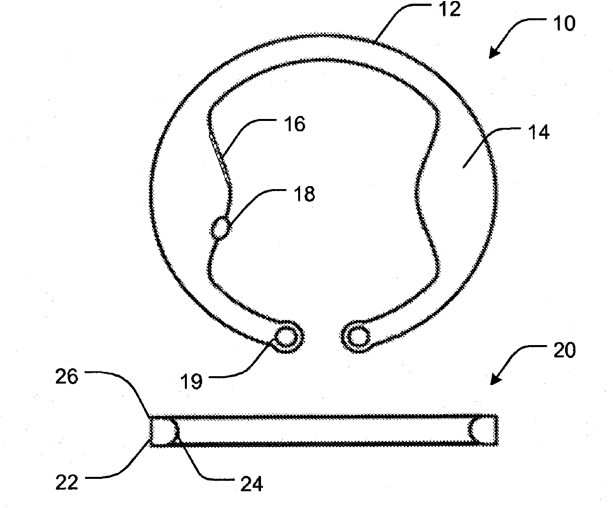
[0048] Standard clear-corneal phacoemulsification was performed on 35 rabbits using intraocular lens (Acrysof SA60AT from Alcon) implants. At each surgery, an intraocular device containing the active agent was inserted into the lens capsule of each rabbit. Rabbits were divided into 4 groups according to the active agent of the intraocular device. The device is loaded with 5-15 mg of Avastin, Timolol, Brimonidine or Latanoprost. Groups were evaluated for stability of intraocular devices and lenses, cystic fibrosis, and healing of cataract wounds and anterior segments. Each eye subgroup was assessed weekly for inflammation for 4 weeks and removed at one month for histopathological assessment of capsule and CDR integrity.
Embodiment 2
[0050] The surgery and procedures described in Example 1 were repeated except that aqueous and vitreous taps were performed every two weeks and utilizing high pressure liquid chromatography (HPLC) and / or enzyme-linked immunosorbent assay (ELISA) Analyze drug concentration. In each drug group, half of the eyes were removed at one month and the other half at two months. This was accomplished by sacrificing the rabbit and enucleating the eye, immediately freezing the eye in liquid nitrogen to prevent perturbation and redistribution of the drug within the ocular tissue. Eyes were then dissected into 3 parts (aqueous humor, vitreous, and retina / choroid layers) to assess anatomic toxicity and tissue drug concentrations. Retrieve the intraocular device and assess the amount of remaining drug. Comparing the Distribution Profile of Intraocular Devices to Conventional Intravitreal Injection of 2.5 mg / 0.1cc Avastin distribution curves for direct comparison of different delivery metho...
Embodiment 3
[0053] Following lens extraction (phacoemulsification technique), three intraocular devices were implanted into eyes of New Zealand white rabbits under general anesthesia. Two devices were loaded with Avastin and one device was loaded with the contrast agent Galbumin as a control. Proper intraocular device placement was verified by magnetic resonance imaging (MRI) and clinical examination.
[0054] One week after implantation the rabbits were sacrificed and the eyes were removed for analysis. The concentration of Avastin in the retina and vitreous was detected by ELISA to be 24-48mcg / mL, and Avastin was present in the eyes of control rabbits. Figure 4 The Avastin content of each eye area analyzed one week after implantation is shown.
PUM
 Abstract
Description
Claims
Application Information
 - Generate Ideas
- Intellectual Property
- Life Sciences
- Materials
- Tech Scout
- Unparalleled Data Quality
- Higher Quality Content
- 60% Fewer Hallucinations
Browse by: Latest US Patents, China's latest patents, Technical Efficacy Thesaurus, Application Domain, Technology Topic, Popular Technical Reports.
© 2025 PatSnap. All rights reserved.Legal|Privacy policy|Modern Slavery Act Transparency Statement|Sitemap|About US| Contact US: help@patsnap.com


