Eureka AIR delivers breakthrough ideas for toughest innovation challenges, trusted by R&D personnel around the world.

Method for evaluating anti-cancer drug inhibiting aggregation of membrane protein receptors

A technology of anti-cancer drug and identification method, which is applied in the field of anti-cancer drug identification, can solve the problems of long cycle, high cost, difficult drug screening, etc., and achieve the effect of short cycle, easy operation, and cost saving

Active Publication Date: 2011-04-27
INST OF CHEM CHINESE ACAD OF SCI
View PDF3 Cites 4 Cited by
  • Summary
  • Abstract
  • Description
  • Claims
  • Application Information

AI Technical Summary

Problems solved by technology

However, in the screening process, there are generally problems of long cycle, complicated operation, and high cost, making it difficult to realize simple and fast drug screening
However, there are no relevant reports on the use of this method to identify anticancer drugs.

Method used


Image

  • Method for evaluating anti-cancer drug inhibiting aggregation of membrane protein receptors
  • Method for evaluating anti-cancer drug inhibiting aggregation of membrane protein receptors
  • Method for evaluating anti-cancer drug inhibiting aggregation of membrane protein receptors

Examples


Embodiment 1

[0032]1) Transfer the prepared HeLa cells to a 35mm glass bottom culture dish and incubate at 37°C for 24 hours to make the cell coverage reach 80%, and then transfect the TβRII-GFP plasmid into this culture dish to make the final concentration Incubate at 37°C for 5 hours in serum-free DMEM at 1 μg / ml to express TβRII-GFP protein on HeLa cells to obtain recombinant cancer cells, then wash the culture dish with serum-free DMEM for 3-5 times.

[0033] The TβRII-GFP plasmid used (Single-molecule imaging reveals transforming growth factor-β-induced type II receptor dimerization; Wei Zhanga, Yaxin Jianga, Qiang Wangb, Xinyong Maa, Zeyu Xiaoa, Wei Zuob, Xiaohong Fanga, 1, and Ye-Guang Chenb, PNAS, September 15, 2009, vol.106, no.37, 15679-15683), the plasmid can be obtained from the Institute of Chemistry, Chinese Academy of Sciences.

[0034] 2) Add naringenin to the cell culture dish used in step 1) to a final concentration of 50 μM, and incubate in serum-free DMEM at 37° C. for ...

Embodiment 2

[0049] According to the same method and steps as in Example 1, the drug to be identified was identified, only the Hela cells used were replaced with MCF7 cells.

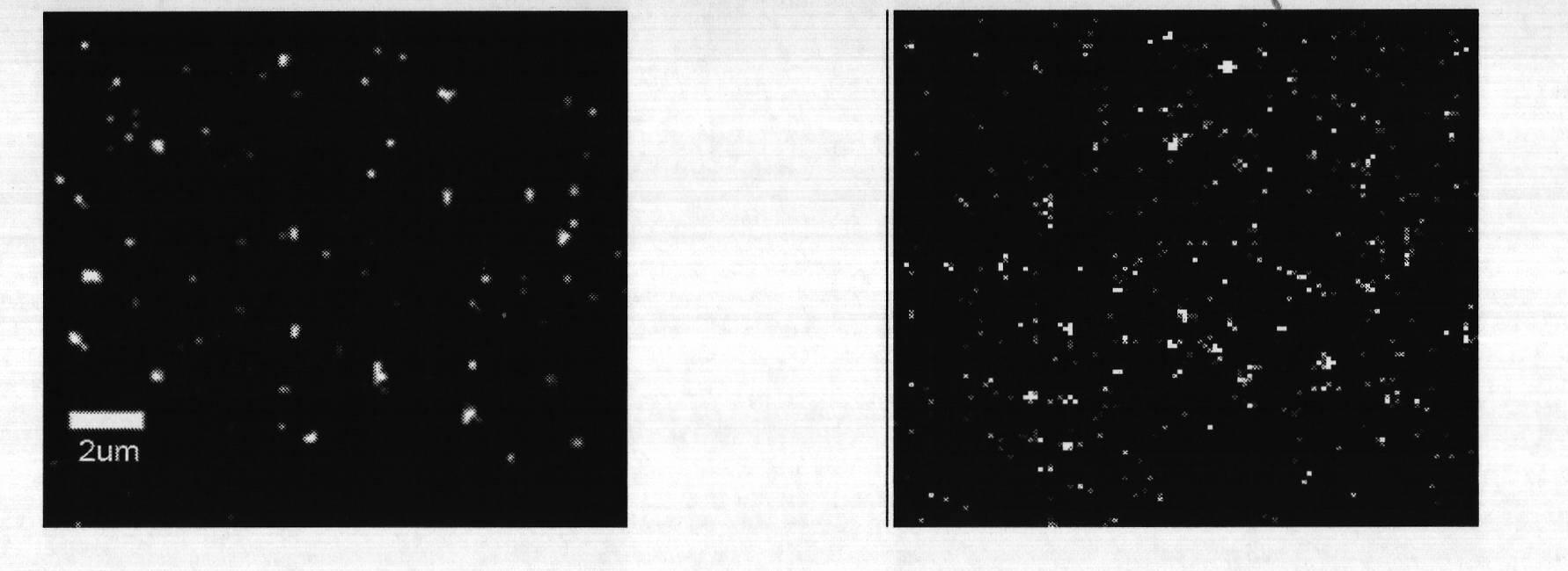
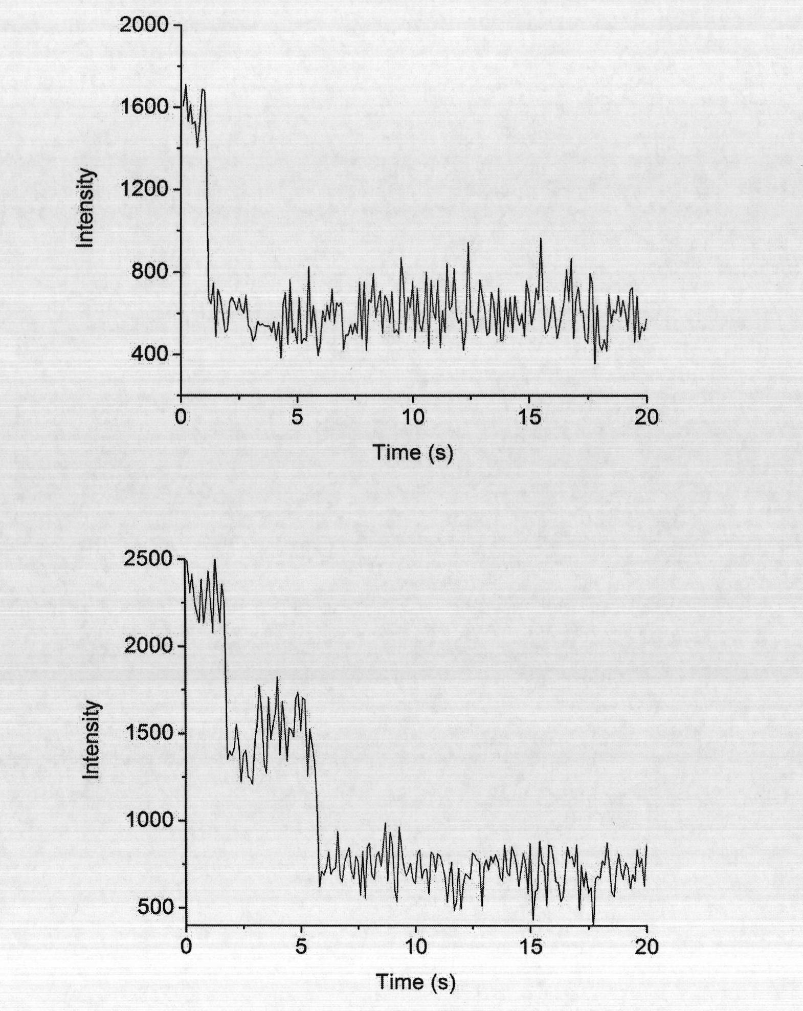
[0050] Fluorescent bright spots less than or equal to 5×5 pixels in the cell images were counted. if so figure 2 The one-step bleaching curve shown on the left is TβRII-GFP monomer. if so figure 2 The two-step bleaching curve shown on the right is TβRII-GFP dimer.

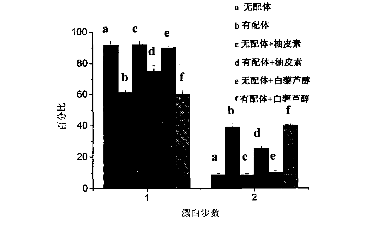
[0051] The results are as follows: the statistics of the bleaching steps of a single fluorescent point are as follows: Figure 4 As shown, when the drug to be identified was not added, after adding the ligand, TβRII-GFP aggregated, and the ratio of TβRII-GFP dimer in the bleaching step A was 37.7±2.9%. After adding 50μM naringenin and incubating for 1h, the proportion of TβRII-GFP aggregation in bleaching step B was significantly reduced, and the proportion of TβRII-GFP dimer decreased to 26.3±1.8%.

[0052] Biochemical experiment verification:

...


PUM

PropertyMeasurementUnit
refractive indexaaaaaaaaaa

Abstract

The invention discloses a method for evaluating an anti-cancer drug inhibiting the aggregation of membrane protein receptors. The method comprises the following steps of: (1) conveying genes of target membrane protein receptors to an excised cancer cell, and marking the genes of the target membrane protein receptors by fluorescent protein to obtain a reconstructed cancer cell; (2) hatching the reconstructed cancer cell and a substance to be evaluated, then hatching by adding ligands of the target membrane protein receptors after the last hatching is finished, and then fixing the reconstructed cancer cell by adding a PBS (Phosphate Buffered Saline) solution of paraformaldehyde to obtain a cell sample I to be detected; and (3) carrying out fluorescence imaging on the unimolecule of the cell sample I to be detected to obtain a cell image and furthermore obtain bleaching steps of each fluorescent spot, and analyzing the image and counting the bleaching steps of the fluorescent spot to judge whether the substance to be evaluated plays an inhibition role in the aggregation of the membrane protein receptors or not. The method has the advantages of simpleness and convenience for operation, short evaluation period and low expense and can be used for evaluating various drugs inhibiting the aggregation of membrane protein receptors.

Description

technical field [0001] The invention relates to an anticancer drug identification method for inhibiting aggregation of membrane protein receptors. Background technique [0002] Cancer has always been the biggest killer of human health. The overproliferation of cells is the cause of the occurrence and development of cancer, and the overproliferation of cells is generally related to the overactivation of ligand-induced cell signaling pathways, especially growth factor signaling pathways. Therefore, inhibiting the excessive activation of this cell signaling pathway has become an important way to treat cancer. The activation of the growth factor cell signaling pathway is firstly activated by the binding of the ligand and its membrane protein receptor on the cell membrane, which causes the receptor to self-polymerize or heteropolymerize. Then, the activated receptor phosphorylates the protein of the downstream pathway, so that the downstream protein is activated, thereby activa...

Claims


Application Information

Patent Timeline
Patent Type & Authority Applications(China)
IPC IPC(8): G01N21/64C12N15/65C12N15/85
Inventor 方晓红杨勇张伟梁伟
Owner INST OF CHEM CHINESE ACAD OF SCI
Why Eureka
Eureka Blog
Learn More