Auxiliary devices and methods for microscopy systems

A technology of auxiliary devices and microscope systems, applied in microscopes, measuring devices, optics, etc., can solve the problems of high price, high use and maintenance costs of flow cytometers, and achieve high cost performance and avoid the trouble of repeated disassembly and installation. Effect

Inactive Publication Date: 2011-12-21
韦宁
View PDF4 Cites 4 Cited by
  • Summary
  • Abstract
  • Description
  • Claims
  • Application Information

AI Technical Summary

Problems solved by technology

However, due to the high price of flow cytometer and the high cost of use and maintenance, its application is limited to a certain extent.

Method used


Image

  • Auxiliary devices and methods for microscopy systems
  • Auxiliary devices and methods for microscopy systems
  • Auxiliary devices and methods for microscopy systems

Examples


Embodiment Construction

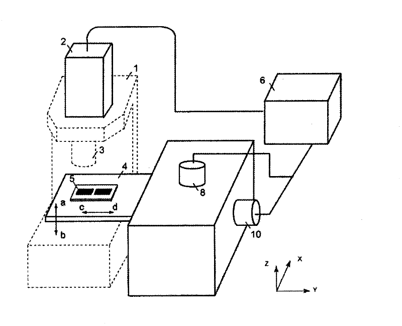
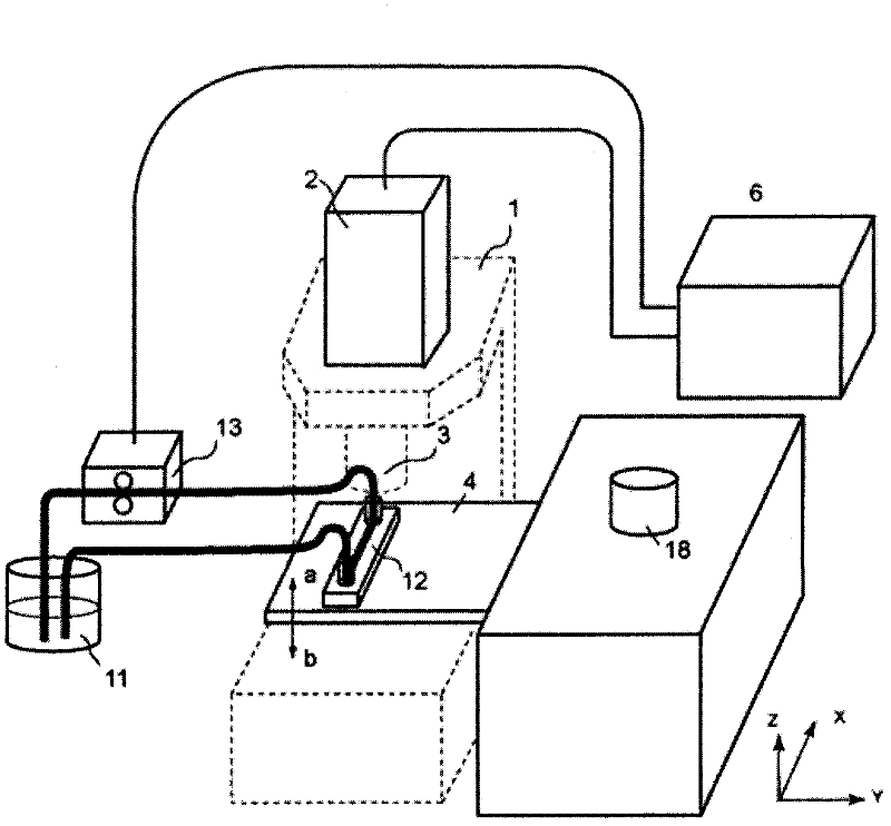
[0078] According to the concept of the present invention, firstly, an auxiliary device independent of the microsystem is placed next to the microsystem. The sample to be tested is then placed on a storage platform. The load-bearing of the object platform is not through the body of the microscope system, but through the auxiliary device itself. A mechanism of the auxiliary device can adjust the relative position between the object holding platform and the microscope system, so that the image acquisition device can acquire images of the sample in different spatial positions. Automatic focusing and sample scanning can be realized through the signal processing unit, and the image of the sample can be processed through the software program to identify, count and classify the particles in the sample.

[0079] According to the invention, this independent device is placed next to the microscope. The sample is then placed on a holder attached to the device. The position of the sampl...


PUM


Abstract

The utility model relates to an auxiliary device for a microscope system, which is independent of the microscope system and comprises a material putting platform for putting a sample, a displacement adjusting mechanism and a signal processing unit. The material putting platform is not carried by the main body of the microscope system. The displacement adjusting mechanism is connected with the material putting platform for adjusting the relative position among the material putting platform, the sample on the material putting platform and the microscope system. The signal processing unit is electrically connected with the displacement adjusting mechanism for controlling at least the partial adjusting act of the displacement adjusting mechanism and controlling an image acquiring device to acquire a sample image. The auxiliary device is easy to integrate into the microscope system so as to cooperatively work therewith and easy to separate from the microscope system so as to avoid the trouble caused by repeated assembly and disassembly.

Description

technical field [0001] The invention relates to an auxiliary device and method for a microscopic system, which can be adapted to microscopic systems of various specifications, can automate, simplify and standardize the analysis process, avoid errors caused by human error, and can expand the display system. Microsystem functions. Background technique [0002] Microscopy, such as bright field, dark field, phase contrast, fluorescence, differential interference, laser confocal microscopy, etc., are widely used to detect and analyze the microstructure of matter. In biological laboratories, microscopic systems or devices are used to observe cells, tiny structures and particles. [0003] When using a microscopic system for analysis, the traditional process is: the experimenter places the collected sample on a carrier (such as a glass slide), and then places the transparent medium on the stage of the microscopic system. Next, the experimenter observes different areas of the sampl...

Claims


Application Information

Patent Timeline
Patent Type & Authority Applications(China)
IPC IPC(8): G02B21/36G02B21/24G02B21/06G01N21/01
CPCG02B21/26
Inventor 韦宁
Owner 韦宁
Why Eureka
Try Eureka