Eureka AIR delivers breakthrough ideas for toughest innovation challenges, trusted by R&D personnel around the world.

Ophthalmology oct system and ophthalmology imaging method

An ophthalmology and scanning imaging technology, which is applied in the field of optoelectronics, can solve the problems that the OCT imaging quality cannot be well matched, and achieve the effect of simple structure, convenient operation, and clear fixation point

Active Publication Date: 2012-05-02
SHENZHEN CERTAINN TECH CO LTD +1
View PDF6 Cites 59 Cited by
  • Summary
  • Abstract
  • Description
  • Claims
  • Application Information

AI Technical Summary

Problems solved by technology

[0006] The technical problem to be solved by the embodiments of the present invention is to provide an ophthalmic OCT system and an ophthalmic OCT imaging method, which can solve the problem that the gaze point adjustment and the OCT imaging quality cannot be well matched in the prior art, and the system structure is simple, easy to operate

Method used


Image

  • Ophthalmology oct system and ophthalmology imaging method
  • Ophthalmology oct system and ophthalmology imaging method
  • Ophthalmology oct system and ophthalmology imaging method

Examples


Embodiment Construction

[0043] The following will clearly and completely describe the technical solutions in the embodiments of the present invention with reference to the accompanying drawings in the embodiments of the present invention. Obviously, the described embodiments are only some, not all, embodiments of the present invention. Based on the embodiments of the present invention, all other embodiments obtained by persons of ordinary skill in the art without creative efforts fall within the protection scope of the present invention.

[0044] Such as image 3 Shown is a schematic block diagram of the first embodiment of the ophthalmic OCT system of the present invention, the ophthalmic OCT system 1 includes a posterior segment OCT imaging system 1a and an anterior segment OCT imaging system 1b, and the posterior segment OCT imaging system 1a also includes an optical path conversion device 110. The anterior segment OCT imaging system 1b includes an anterior segment probe imaging device 1b1, and th...


PUM


Abstract

The embodiment of the invention discloses an ophthalmology OCT system, comprising a posterior segment intraocular OCT imaging system and an anterior eye segment OCT imaging system. The posterior segment intraocular OCT imaging system comprises a light path conversion device, the anterior eye segment OCT imaging system comprises an anterior eye segment probe imaging device comprising an ophthalmoscope, a first dichroscope and a first lens; wherein, when the ophthalmology OCT system carries out an anterior eye segment scanning imaging, and after the light path conversion device receives a light path conversion instruction, signal light passing through a collimating mirror and a scanning device respectively are converted to the anterior eye segment probe imaging device; the anterior eye segment probe imaging device drives the first dichroscope to reflect the signal light to the ophthalmoscope to carry out the anterior eye segment scanning imaging. Correspondingly, the invention also discloses an ophthalmology OCT imaging method. By employing the invention, when the anterior eye segment imaging is carried out on human eye, fixation points can always be kept clear via refractive compensation, and the anterior eye segment probe imaging quality is not affected.

Description

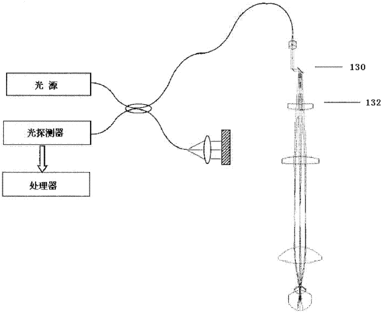
technical field [0001] The invention relates to the field of optoelectronics, in particular to an ophthalmic OCT system and an ophthalmic OCT imaging method. Background technique [0002] Optical coherence tomography (OCT, Optical Coherence Tomography) is an emerging optical imaging technology. Compared with traditional clinical imaging methods, it has the advantages of high resolution, imaging speed, no radiation damage, moderate price, and compact structure. , is an important potential tool for basic medical research and clinical diagnostic applications. Currently, among various ophthalmic devices using optical instruments, an OCT device for ophthalmic examination and treatment has become an indispensable ophthalmic device for ophthalmic disease diagnosis. [0003] Patent document US2009 / 0168017A1 discloses an OCT device for realizing anterior segment imaging in a posterior segment OCT system, such as figure 1 As shown, a lens 132 is inserted behind the scanning galvanom...

Claims


Application Information

Patent Timeline
Patent Type & Authority Applications(China)
IPC IPC(8): A61B3/14
CPCA61B3/102G01N21/4795
Inventor 蔡守东郭曙光代祥松李鹏吴蕾
Owner SHENZHEN CERTAINN TECH CO LTD
Why Eureka
Eureka Blog
Learn More