In vitro amplification and low-temperature storage method for regulatory T cells of umbilical cord blood

A technology for cryopreservation and umbilical cord blood, applied in the field of cell expansion and preservation in vitro

Active Publication Date: 2012-06-27
SHANGHAI BLOOD CENT
View PDF3 Cites 18 Cited by
  • Summary
  • Abstract
  • Description
  • Claims
  • Application Information

AI Technical Summary

Problems solved by technology

However, there is no report about a method for in vitro expansion and cryopreservation of umbilical cord blood regulatory T cells.

Method used


Image

  • In vitro amplification and low-temperature storage method for regulatory T cells of umbilical cord blood
  • In vitro amplification and low-temperature storage method for regulatory T cells of umbilical cord blood
  • In vitro amplification and low-temperature storage method for regulatory T cells of umbilical cord blood

Examples


Embodiment 1

[0034] Add 6% hydroxyethyl starch substitute slurry to cord blood at a volume ratio of 4:1, place in a refrigerator at 4°C for 1 hour to precipitate red blood cells, take the upper plasma layer and centrifuge (2000rpm / min, 5min), discard most of the plasma, and precipitate white blood cells. After suspension, they were separated with Ficoll lymphocyte separation medium (2000rpm / min, 20min), and washed three times with PBS to obtain umbilical cord blood mononuclear cells. According to the manufacturer's instructions, use standard separation kits, such as CliniMACS, Miltenyi Biotec's anti-human CD8, CD14, CD19, CD56 monoclonal antibody immunomagnetic beads to remove non-CD4 cells in the cord blood mononuclear cells, and then isolate the cord blood CD4+ T cells Enrich CD4+CD25+ cells with anti-human CD25 antibody-coupled magnetic beads. After the cord blood CD4+CD25+T cells were purified, the purity of the isolated cells was detected by flow cytometry.

Embodiment 2

[0036] In vitro expansion of purified cord blood CD4+CD25+ T cells with anti-human CD3 / CD28 antibody-coupled magnetic beads and recombinant human IL-2 (200 U / ml) in commercially available cell culture bags or cell culture plates , using X-VIVO15 complete culture medium (containing 10% inactivated human AB serum, penicillin and streptomycin double antibody 50U / ml), the ratio of CD3 / CD28-coated Dynal Beads to cells in the culture system is 1:1 , at 37℃, 5%CO 2 Cultured under the conditions of humidity and saturated humidity, half of the culture medium and IL-2 were replaced every other day. The whole expansion time is 2-3 weeks, including two rounds of expansion of 6-8 days each and two rounds of expansion in the middle of which the cells rest for 2-4 days, that is, after the cells are removed from the magnetic beads, the cells contain IL-2 (20U / ml ) of X-VIVO15 complete culture medium for 2 days. After the in vitro expansion, the number of cells increased by about 1500 times,...

Embodiment 3

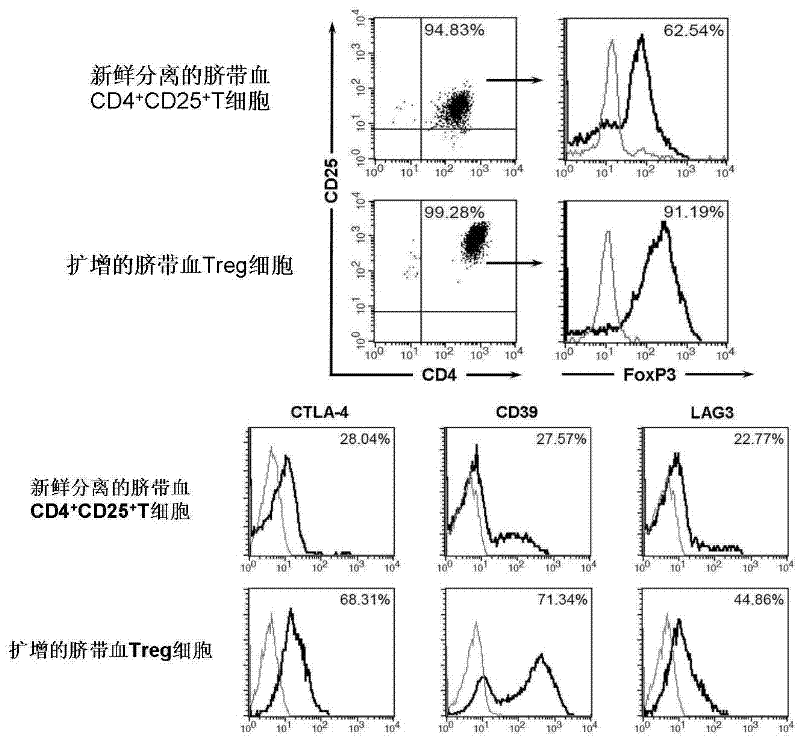
[0038] Immunophenotyping of cord blood Treg cells was performed by flow cytometry. Use FITC-labeled CD4 antibody, PE-Cy5-labeled CD25 antibody, APC-labeled CD127 antibody, PE-labeled CTLA-4 antibody, CD39 antibody, and LAG3 antibody for cell surface labeling. PE-tagged Foxp3 for intracellular labeling. Such as figure 1 As shown, the expanded cord blood Treg cells co-expressed CD4 and CD25, and more than 90% of the cells expressed FoxP3. At the same time, with freshly sorted CD4 + CD25 + Compared with T cells, expanded cord blood Treg cells highly expressed CTLA-4 (68%), CD39 (71%) and LAG-3 (45%).


PUM


Abstract

The invention relates to an in vitro amplification and low-temperature storage method for regulatory T cells of umbilical cord blood. The method comprises the following steps of: separating a CD4+CD25+T cell group rich in the regulatory T cells from the umbilical cord blood; amplifying the enriched CD4+CD25+T cell group to obtain high-purity regulatory T cells; and storing the amplified regulatory T cells in liquid nitrogen at a low temperature to serve as third-party unrelated donor regulatory T cells for treating clinical diseases. The method has the advantages that: a large quantity of regulatory T cells can be obtained from the umbilical cord blood and stored for a long term in the liquid nitrogen, can meet the requirements of clinic for regulatory T cell products, and are used for treating graft versus host disease, organ transplantation rejection and autoimmune disease.

Description

technical field [0001] The invention relates to a method for expanding and preserving cells in vitro, in particular to a method for expanding and storing umbilical cord blood regulatory T cells in vitro and at low temperature. Background technique [0002] Rejection of MHC-mismatched grafts by recipients is a major cause of tissue and cell transplantation failure. In current clinical practice, although the use of traditional immunosuppressive drugs can effectively prevent transplant rejection and alleviate acute and chronic graft-versus-host disease (GVHD) mediated by donor T cells, it is often Accompanied by the decline of the recipient's overall immune function, increased chances of infection and induced tumors and other side effects. Therefore, cell therapy methods aimed at inducing immune tolerance of transplantation between donors and recipients have been paid more and more attention. CD4+CD25+Foxp3+regulatory T cells (regulatory T cells, Tregs), as a T cell subset wi...

Claims


Application Information

Patent Timeline
Patent Type & Authority Applications(China)
IPC IPC(8): C12N5/0783A01N1/02A61K35/44A61K35/14A61P37/02A61P37/06A61K35/51
Inventor 范华骅杨洁杨懿铭任亚娜谢如锋
Owner SHANGHAI BLOOD CENT
Why Eureka
Try Eureka