Fluorescent endoscopic imaging method and system

An imaging method and imaging system technology, applied in the fields of endoscopy, medical science, diagnosis, etc., can solve the problems of low efficiency and slow fluorescence endoscopic imaging, and achieve the effects of fast speed, little damage to organisms, and short time.

Inactive Publication Date: 2012-07-04
SHENZHEN UNIV
View PDF4 Cites 4 Cited by
  • Summary
  • Abstract
  • Description
  • Claims
  • Application Information

AI Technical Summary

Problems solved by technology

[0004] The purpose of the embodiments of the present invention is to provide a fluorescent endoscopic imaging method, aiming to solve the problems of slow and low efficiency of existing fluorescent endoscopic imaging

Method used


Image

  • Fluorescent endoscopic imaging method and system
  • Fluorescent endoscopic imaging method and system
  • Fluorescent endoscopic imaging method and system

Examples


Embodiment Construction

[0024] In order to make the object, technical solution and advantages of the present invention clearer, the present invention will be further described in detail below in conjunction with the accompanying drawings and embodiments. It should be understood that the specific embodiments described here are only used to explain the present invention, not to limit the present invention.

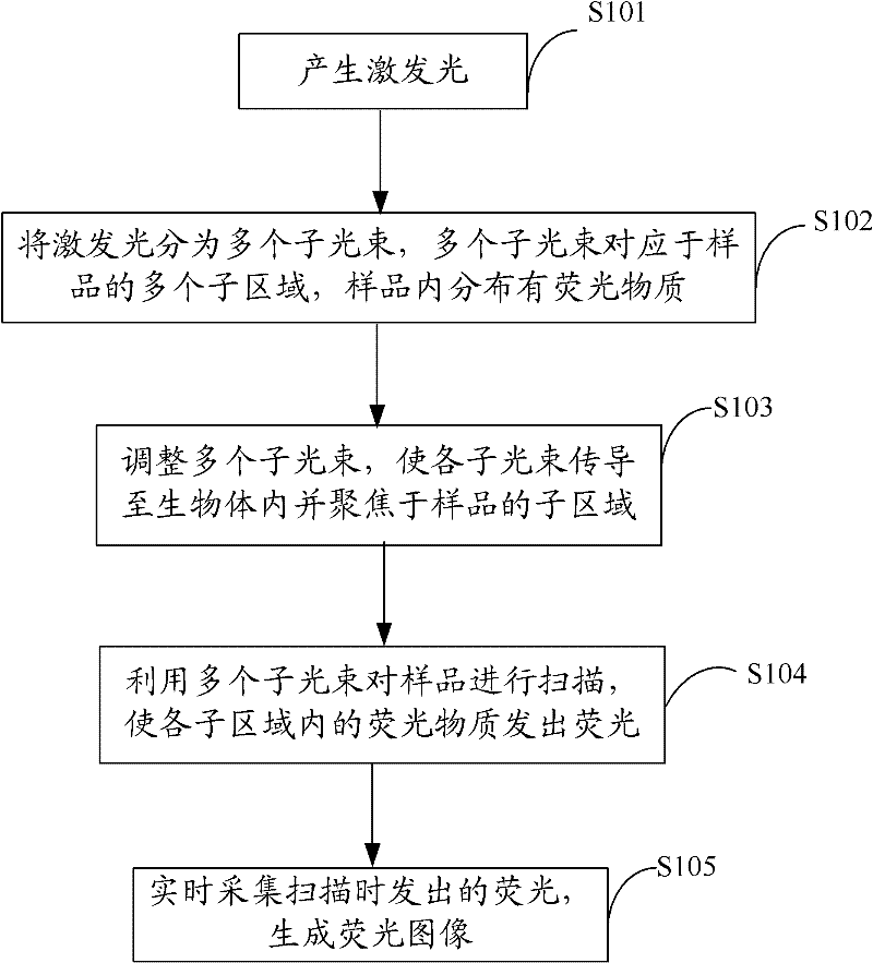
[0025] In the embodiment of the present invention, the excitation light is divided into multiple sub-beams corresponding to multiple sub-regions of the sample, and the multiple sub-beams are transmitted into the living body, and each sub-beam is focused on a sub-region of the sample to form a multi-point excitation fluorescence, and the fluorescence It is exported, and the sample is scanned two-dimensionally by multiple sub-beams, so as to obtain the fluorescence image of the entire sample, which is short in time, fast in speed, and has little damage to organisms, which is beneficial to biomedical r...


PUM


Abstract

The invention is applicable to the filed of photoelectric detection, and provides a fluorescent endoscopic imaging method and a fluorescent endoscopic imaging system, wherein the fluorescent endoscopic imaging method comprises the following steps: generating exciting light; dividing the exciting light into a plurality of sub light beams which correspond to a plurality of subareas of a sample, wherein a fluorescent material is distributed in the sample; adjusting the plurality of sub light beams till the sub light beams are conducted to an organism and are focused on the subareas of the sample; utilizing the sub light beams to scan the sample till the fluorescent material in the subareas emits fluorescence; and collecting the fluorescence generated in scanning in real time to generate a fluorescent image. For the florescent endoscopic imaging method, the plurality of sub light beams are utilized for carrying out two-dimensional scanning on the sample, so that the fluorescent image of the whole sample is acquired, the time consumed is short, the speed is fast, the harm to the organism is small, the research on biomedicine is facilitated, and the fluorescent endoscopic imaging method and the fluorescent endoscopic imaging system have great significance for early diagnosis of cancer.

Description

technical field [0001] The invention belongs to the field of photoelectric detection, in particular to a fluorescent endoscopic imaging method and system. Background technique [0002] Fluorescence microscopy has become an important tool in life sciences, especially cell biology research. Multi-photon excitation fluorescence microscopy has the advantages of small killing effect on living organisms, large penetration depth, and chromatographic ability, and has become an important means of life science research. Fluorescence images can provide structural and functional information for biomedical detection and analysis. [0003] In recent years, with the rapid development of new optical fiber and micro-manufacturing technology, the research of fiber-optic two-photon fluorescence microscope and endoscope has made it possible to study two-photon fluorescence microscopy imaging technology in living internal organs and living animals. At present, two-photon fluorescence endoscopi...

Claims


Application Information

Patent Timeline
Patent Type & Authority Applications(China)
IPC IPC(8): A61B5/00
CPCA61B1/07A61B1/043A61B1/00172
Inventor 邵永红屈军乐牛憨笨
Owner SHENZHEN UNIV
Why Eureka
Try Eureka