Method for preparing scanning electron microscope samples from biological samples
A biological sample, scanning electron microscope technology, applied in the preparation of test samples, etc., can solve the problems of low accuracy, different first protein and second protein, etc.
- Summary
- Abstract
- Description
- Claims
- Application Information
AI Technical Summary
Problems solved by technology
Method used
Image
Examples
Embodiment approach
[0026] According to a preferred embodiment of the method according to the invention, said first contacting is carried out in the presence of bovine serum albumin at a concentration of 0.5-2% by weight.
[0027] According to the method of the present invention, wherein, the conditions of the second contact are not particularly required, and can be conventional conditions of immunoelectron microscopy, for example (The Journal of Cell Biology (The Journal of Cell Biology), 1984, volume 99, page 53 ) conditions described in the literature, in order to make the prepared scanning electron microscope sample can accurately distinguish two different protein molecules, preferably, the conditions of the second contact include: relative to each mg of biological sample, labeled with The amount of the first antibody of the first gold nanoparticle or the second antibody labeled with the second gold nanoparticle is 100-800 micrograms, more preferably 200-600 micrograms; the contact temperature...
preparation example 1
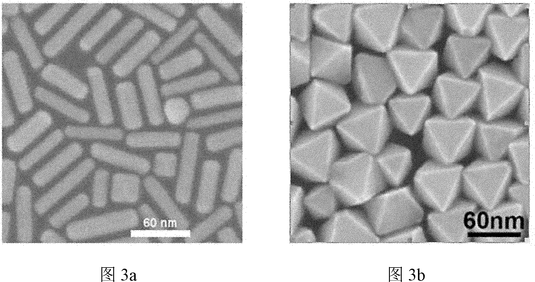
[0063] This preparation example is used to illustrate the method for preparing the first antibody labeled with the first gold nanoparticles and the second antibody labeled with the second gold nanoparticles.
[0064] 1. Prepare the following solution:
[0065] (1) 0.1M cetyltrimethylammonium bromide (C 19 h 42 N + Br - , hereinafter referred to as CTAB) aqueous solution;
[0066] Weigh 3.64g of cetyltrimethylammonium bromide (analytical grade) and dissolve in 100mL of deionized water; before use, it should be placed in a constant temperature water bath at 30°C to dissolve completely to obtain 0.1M cetyltrimethylammonium bromide ammonium bromide (C 19 h 42 N + Br - , hereinafter referred to as CTAB) aqueous solution;
[0067] (2) 0.1M aqueous ascorbic acid (AA) aqueous solution:
[0068]Weigh 0.176g of ascorbic acid (analytical pure) and dissolve it in 10mL of deionized water to obtain a 0.1M ascorbic acid (AA) aqueous solution (the solution can be temporarily prepare...
preparation example 2
[0099] This preparation example is used to illustrate the preparation method of the biological material mentioned in the present invention.
[0100] The preparation purpose of this preparation example is to prepare rat brain microvascular endothelial cells stimulated by burn serum, and the steps are as follows:
[0101] (1) Primary culture of rat brain microvascular endothelial cells:
[0102] Adult Wistar male rats were anesthetized with 3.5% by weight of chloral hydrate and then decapitated to take out the brain tissue, which was placed in a petri dish; the pia mater, large blood vessels and white matter were removed to obtain the cerebral cortex, and the obtained cerebral cortex was cut After crushing, use Dounce (Dounce) homogenizer to homogenate to obtain a homogenate, and filter the homogenate with a 180-mesh filter to obtain a filtrate; Digest at 37°C for 15 minutes to obtain a digestive solution; centrifuge the digestive solution at a speed of 800g for 5 minutes and d...
PUM
| Property | Measurement | Unit |
|---|---|---|
| diameter | aaaaa | aaaaa |
| length | aaaaa | aaaaa |
| diameter | aaaaa | aaaaa |
Abstract
Description
Claims
Application Information
 - R&D
- Intellectual Property
- Life Sciences
- Materials
- Tech Scout
- Unparalleled Data Quality
- Higher Quality Content
- 60% Fewer Hallucinations
Browse by: Latest US Patents, China's latest patents, Technical Efficacy Thesaurus, Application Domain, Technology Topic, Popular Technical Reports.
© 2025 PatSnap. All rights reserved.Legal|Privacy policy|Modern Slavery Act Transparency Statement|Sitemap|About US| Contact US: help@patsnap.com


