Preservation method for corneal limbus tissue

A preservation method and corneal limbal technology, applied in the preservation, application, animal husbandry and other directions of human or animal body, can solve the problems of loss of epithelium, damage to the basement membrane, limiting the clinical application of limbal active tissue, etc. Active, simple production process effect

Active Publication Date: 2012-10-17
XIAMEN EYE CENTER OF XIAMEN UNIVERSITY CO LTD
View PDF10 Cites 6 Cited by
  • Summary
  • Abstract
  • Description
  • Claims
  • Application Information

AI Technical Summary

Problems solved by technology

During storage, traditional corneal materials will lose epithelium and even damage the basement membrane, which limits the clinical application of limbal active tissue to a certain extent.

Method used


Image

  • Preservation method for corneal limbus tissue
  • Preservation method for corneal limbus tissue
  • Preservation method for corneal limbus tissue

Examples


Embodiment 1

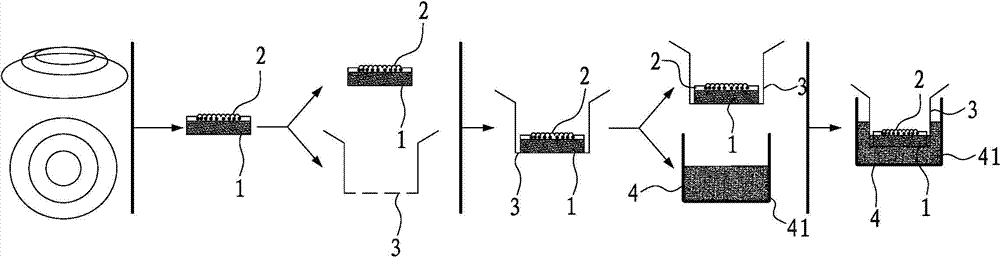
[0045] The preparation method of the present invention comprises the following steps: the residual corneal limbal tissue after the corneal transplantation of the dead eyeball is cleaned with sterile physiological saline in a sterile and clean environment, and then disinfected. Fresh corneal limbal tissue is soaked and rinsed with antibiotic-containing phosphate buffered saline for 5-10 minutes, rinsed at least 3 times, each time greater than or equal to 5 minutes; the operator wears sterile gloves on both hands, and removes the conjunctiva and iris with microscopic instruments tissue, put the sterile transwell chamber in a sterile cell culture dish with a diameter of 35mm, spread the corneal limbal tissue on the PVDF membrane of the transwell chamber; Contact; place the cell culture dish in the refrigerator to keep the ambient temperature constant at 4°C, and replace with fresh sterile medium-term preservation solution every 2 days. In this embodiment, the PVDF film is a speci...

Embodiment 2

[0047] The preparation method of the present invention comprises the following steps: the residual corneal limbal tissue after the corneal transplantation of the dead eyeball is cleaned with sterile physiological saline in a sterile and clean environment, and then disinfected. Fresh corneal limbal tissue is soaked and rinsed with antibiotic-containing phosphate buffered saline for 5-10 minutes, rinsed at least 3 times, each time greater than or equal to 5 minutes; the operator wears sterile gloves on both hands, and removes the conjunctiva and iris with microscopic instruments tissue, put the sterile transwell chamber in a sterile cell culture dish with a diameter of 35mm, spread the corneal limbal tissue on the PVDF membrane of the transwell chamber; Contact; place the cell culture dish in an incubator, keep the ambient temperature constant at 37°C, and replace with fresh sterile medium-term preservation solution every 2 days. In this embodiment, the PVDF film is a specific i...

Embodiment 3

[0049] The preparation method of the present invention comprises the following steps: the residual corneal limbal tissue after the corneal transplantation of the cadaveric source eyeball is washed with sterile physiological saline to clean the blood stains on the surface, and then disinfected. Fresh corneal limbal tissue is soaked and rinsed with antibiotic-containing phosphate buffered saline for 5-10 minutes, rinsed at least 3 times, each time greater than or equal to 5 minutes; the operator wears sterile gloves on both hands, and removes the conjunctiva and iris with microscopic instruments tissue, put the sterile transwell chamber in a sterile cell culture dish with a diameter of 35 mm, spread the corneal limbal tissue on the PVDF membrane of the transwell chamber; add SHEM medium at the bottom of the transwell chamber until the liquid level is fully covered with the PVDF membrane Contact; place the cell culture dish in a refrigerator to keep the ambient temperature constan...


PUM


Abstract

The invention discloses a preservation method for corneal limbus tissue. The method comprises a step of disinfecting fresh corneal limbus tissue and a step of air exposure preservation of the disinfected fresh corneal limbus tissue. The step of air exposure preservation is as follows: the fresh corneal limbus tissue is placed on a permeable covering film, preservation liquid or a cell culture medium capable of providing nutrients to the corneal limbus tissue is arranged below the covering film, epitheliums of cornea directly contact with air, the temperature of a culture environment in the step is 0 to 37 DEG C, and oxygen concentration is 0 to 20%. Compared to immersion preservation in the prior art, the preservation method provided in the invention has the following advantages: the structure of the preserved corneal limbus tissue is more integral; obvious apoptosis of the epitheliums of cornea does not occur; stem cells of corneal limbus can maintain high activity; and process flow is simple and reliable, is easy to implement, costs little and has other efficacy.

Description

technical field [0001] The invention relates to the field of preservation of transplanted materials, in particular to a method for preservation of limbal tissue. Background technique [0002] Limbal tissue is widely used in many diseases such as limbal stem cell deficiency or dysfunction, limbal corneal degeneration and limbal tumors. One of the important issues in this treatment is the removal of the limbal tissue from the donor and keeping it alive. [0003] In 1992, Kaufman reported the Optisol corneal active preservation solution, which made it safe and reliable to store cornea slices in eye banks within 2-3 weeks, and became the preferred preservation solution for penetrating keratoplasty in eye banks around the world. In China, the 2-4° wet room preservation method is mainly used. Penetrating keratoplasty (PKP) is the mainstream method of traditional keratoplasty. In order to meet the needs of penetrating keratoplasty, corneal grafts focus on providing corneal active...

Claims


Application Information

Patent Timeline
Patent Type & Authority Applications(China)
IPC IPC(8): A01N1/02
Inventor 吴护平董诺李程刘祖国
Owner XIAMEN EYE CENTER OF XIAMEN UNIVERSITY CO LTD
Why Eureka
Try Eureka