Transesophageal ultrasonic visual simulation system and method used for teaching and clinical skill training

A simulation system and transesophageal technology, applied in teaching models, educational tools, instruments, etc., can solve problems such as inaccessibility, slippage, and difficulty in further improving clinical surgical assistance, and achieve the effect of improving accuracy and prolonging service life

Active Publication Date: 2014-08-06
WEST CHINA HOSPITAL SICHUAN UNIV +1
View PDF8 Cites 0 Cited by
  • Summary
  • Abstract
  • Description
  • Claims
  • Application Information

AI Technical Summary

Problems solved by technology

However, because the above-mentioned product belongs to a closed teaching system, clinical experience is not easy to be accumulated and expanded, which is not conducive to data sharing between teaching and clinical practice, and it is not easy to add clinical experience to the teaching system at any time
[0006] Japan’s transesophageal ultrasound simulation teaching equipment was successfully developed by Japan’s HRS Medical Systems Inc in 2008. This product includes analog probes, simulators, and image databases, but it does not realize three-dimensional visualization, and the key to TEE learning is two-dimensional ultrasound. The embodiment of the cut surface on the 3D visualized heart is difficult to be further improved to clinical surgical assistance due to the lack of open modeling
[0007] The invention patent of ZL 201010251812.5 "transesophageal echocardiography visualization simulation system and method" realizes the one-to-one matching between the posture data when collecting the transesophageal ultrasound section and the three-dimensional visualization heart section, proposes the pathological mining of heart diseases, and gives the corresponding The spatial position relationship between the ultrasound section and the virtual 3D heart model, but the 3D heart model reconstructed by this product lacks the analysis of the biomechanics and other characteristics of medical tissues, and cannot fully express the true texture of the heart
In addition, due to the unstable physical contact and communication line contact of the magnetic linkage sensor used in this product, when measuring the displacement distance, there will be no contact and slippage, which will cause some echocardiographic probe attitude parameters ( Especially the displacement distance parameter) is not accurate, which greatly reduces the realism and accuracy of the simulation

Method used


Image

  • Transesophageal ultrasonic visual simulation system and method used for teaching and clinical skill training
  • Transesophageal ultrasonic visual simulation system and method used for teaching and clinical skill training
  • Transesophageal ultrasonic visual simulation system and method used for teaching and clinical skill training

Examples


Embodiment 1

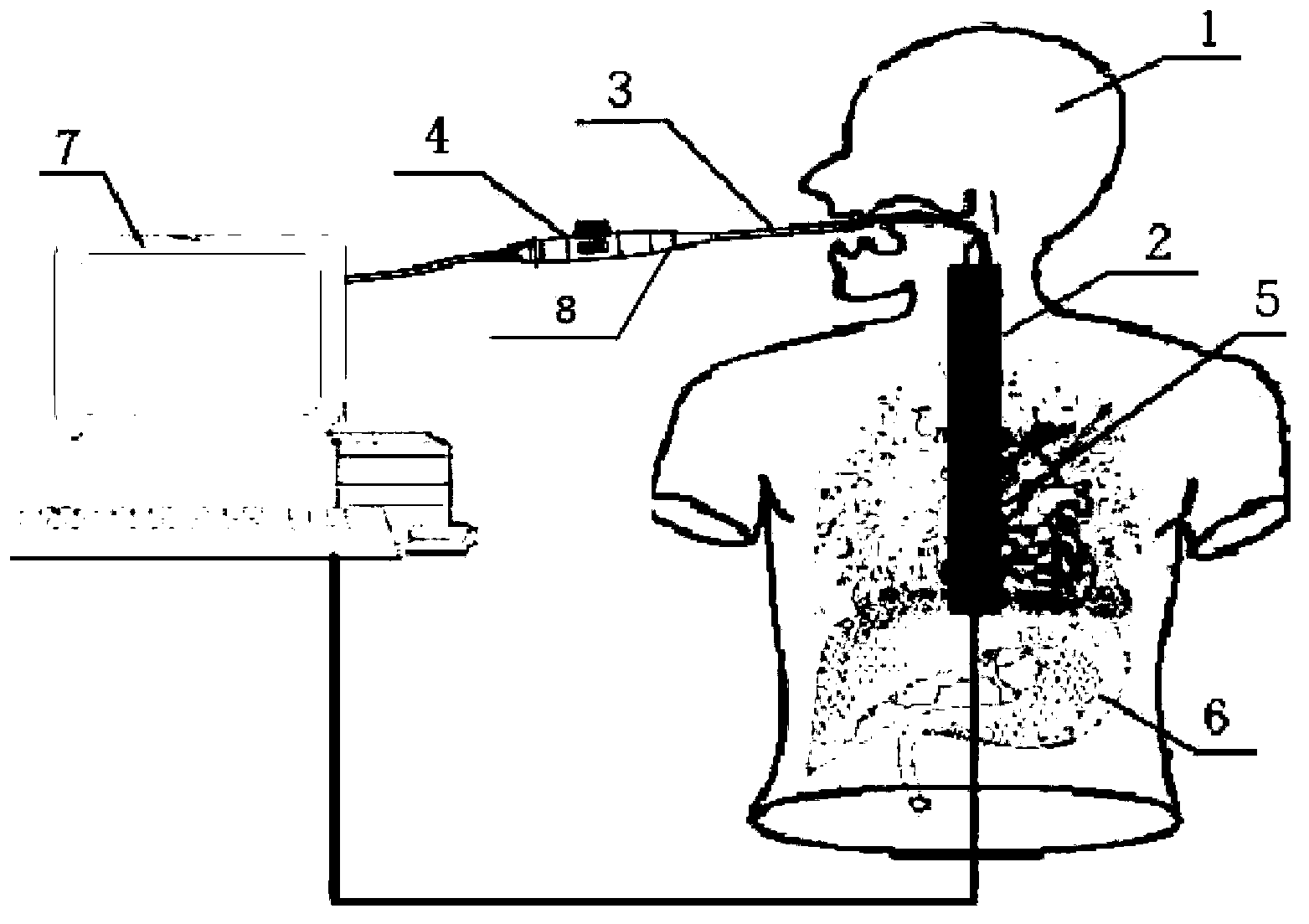
[0031] In this embodiment, the structure of the transesophageal ultrasound visualization simulation system used for teaching and clinical skills training is as follows: figure 1 , figure 2 As shown, it consists of an intelligent phantom 1, a TEE simulation probe 3, a probe attitude device and a computer 7.

[0032] The smart phantom 1 is mainly composed of a phantom and a three-dimensional simulated heart 5, a simulated esophagus and a stomach 6 arranged inside the phantom. The phantom of the smart phantom is made of flesh-colored soft plastic to simulate skin, and the oral cavity of the head can move and can Opens and closes, the head and neck can be separated to allow the rangefinder to be easily placed in the esophagus of the smart phantom.

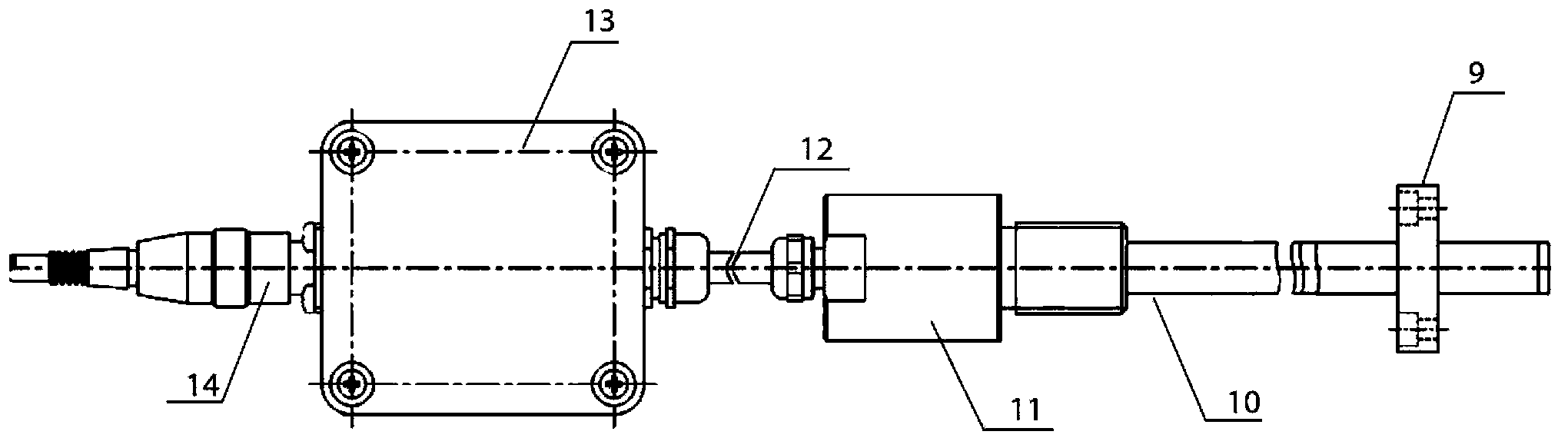
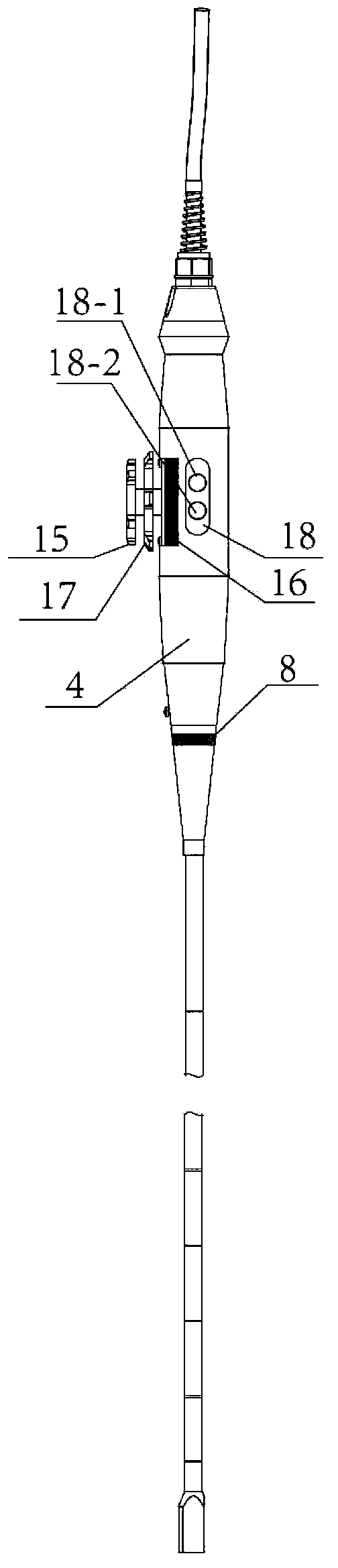
[0033] The probe attitude device is composed of a range finder 2, an angle measuring device 8, a swing simulator and an ultrasonic sector rotation simulator.

[0034] The structure of range finder 2 is as follows figure 2 As shown...

Embodiment 2

[0041]The transesophageal ultrasound visualization simulation method used in teaching and clinical skills training in this embodiment uses the transesophageal ultrasound visualization simulation system described in Example 1, and the steps are as follows:

[0042] The first step is to store the echocardiographic data obtained from the database of clinical ultrasonography instruments in the hospital, and the corresponding relationship data between the probe posture data positioned by the clinical ultrasonography instrument and the transesophageal echocardiogram in the transesophageal echocardiogram described in Example 1. In the computer 7 of the ultrasound visualization simulation system.

[0043] The second step is to build a virtual three-dimensional dynamic heart model and store it in the computer 7 of the transesophageal ultrasound visualization simulation system described in Embodiment 1. The virtual three-dimensional dynamic heart model uses a tree structure to map the he...


PUM


Abstract

The invention relates to a transesophageal ultrasonic visual simulation system used for teaching and clinical skill training. The system comprises an intelligent body model, a TEE (transesophageal echocardiography) simulated probe, a probe posture device and a computer, wherein the probe posture device consists of a diastimeter, an angle measurer, a hook swing simulator and an ultrasonic sector rotation simulator. The invention also relates to a transesophageal ultrasonic visual simulation method, which comprises the following steps: 1, echocardiography data acquired from the data base of a clinical ultrasonograph of a hospital and data of corresponding relation between probe posture data positioned by the clinical ultrasonograph and TEE are stored in the computer of the transesophageal ultrasonic visual simulation system; 2, a virtual three-dimensional dynamic heart model is established and stored in the computer of the transesophageal ultrasonic visual simulation system; and 3, the transesophageal ultrasonic visual simulation system is used in simulated training.

Description

technical field [0001] The invention belongs to the technical field of medical image analysis and virtual simulation, and relates to a transesophageal ultrasound visualization simulation system and method for teaching and clinical skills training. Background technique [0002] Heart disease is the number one killer of human health. 1 / 3 of the world's population death is caused by heart disease, and hundreds of thousands of people die of heart disease every year in my country. Ultrasound diagnosis of heart disease has always been the focus and difficulty in the field of ultrasound diagnosis. Transesophageal echocardiography is a new development in the field of cardiovascular disease diagnosis and monitoring, and provides a new means for the diagnosis of various heart diseases and large vessel diseases. The structure and movement of the heart are very complex, which puts forward higher requirements for the examiner's spatial thinking ability and clinical thinking ability. Rese...

Claims


Application Information

Patent Timeline
Patent Type & Authority Patents(China)
IPC IPC(8): G09B23/28
Inventor 宋海波苗青刘进陈晓清王伟东雷超付忠良姚宇刘亮亮唐红张丹普
Owner WEST CHINA HOSPITAL SICHUAN UNIV
Why Eureka
Try Eureka