Inducing method for directionally differentiating human embryonic stem cells to corneal endothelial cells

A technology of human embryonic stem cells and corneal endothelium, applied in the field of tissue engineering and ophthalmology, can solve the problems of inability to obtain human neural crest stem cells and hinder the induction and differentiation of human corneal endothelial cells

Active Publication Date: 2013-03-06
SHANDONG UNIV
View PDF0 Cites 24 Cited by
  • Summary
  • Abstract
  • Description
  • Claims
  • Application Information

AI Technical Summary

Problems solved by technology

Involving ethical controversy, we have not been able to obtain human neural crest stem cells through primary culture, which hinders the induction of human corneal endothelial cell differentiation

Method used


Image

  • Inducing method for directionally differentiating human embryonic stem cells to corneal endothelial cells
  • Inducing method for directionally differentiating human embryonic stem cells to corneal endothelial cells
  • Inducing method for directionally differentiating human embryonic stem cells to corneal endothelial cells

Examples


Embodiment

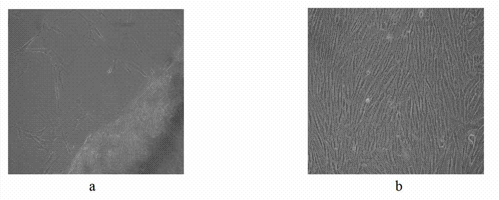
[0028] 1. Primary culture of human corneal stromal fibroblasts: obtain fresh donor corneal rings that are not clinically suitable for corneal transplantation, soak them in balanced salt solution containing 100U / ml penicillin-100U / ml streptomycin repeatedly Rinse 3 times, 10 minutes each time, place under an inverted microscope, scrape off the corneal epithelium with an epidermic knife, tear off the elastic layer and endothelium with microscopic tweezers, and rinse the remaining corneal stroma repeatedly with balanced salt solution to remove residual corneal epithelium and endothelial fragments , cut it into 1mm×1mm tissue pieces with micro scissors, and evenly adhere to the culture dish. After the wall is firmly attached, add DMEM / F12 medium containing 10% fetal bovine serum, and place at 37°C, 5%CO 2 Incubation in the incubator. The medium was changed every 3 days, and some stromal cells were found to swim out after 7 days of culture (such as figure 1 As shown in a), after a...


PUM


Abstract

The invention discloses an inducing method for directionally differentiating human embryonic stem cells to corneal endothelial cells. The method comprises the steps of: cultivating the human embryonic stem cells on a mouse embryonic fibroblast feed layer; sorting human embryonic stem cell clone groups in good state; grafting the groups on a human corneal stromal fibroblast layer processed by mitomycin C and cultivating for 7 days, wherein the human embryonic stem cells are differentiated to rosettes; separating and transferring the rosettes from the human corneal stromal fibroblast layer to a culture bottle; cultivating continuously for 7 days by using a neural crest stem cell culture medium; sorting the neural crest stem cells by a flow cytometry; adding the neural crest stem cells into the culture bottle; placing in a 5% CO2 incubator for incubating and cultivating at 37 DEG C by using a human corneal endothelial cell culture medium; changing the liquid every other day; and cultivating for about 10 days to obtain the corneal endothelial cells. The multiplication capacity of the corneal endothelial cells are similar to that of human corneal endothelial cells and the corneal endothelial cells can be transferred to 1-2 generations in vitro maximally. The corneal endothelial cells can be used as seed cells for cornea construction and transplant in tissue engineering.

Description

technical field [0001] The invention relates to an induction method for directional differentiation of human embryonic stem cells into corneal endothelial cells, and use of the corneal endothelial cells obtained by the induction method as seed cells for tissue engineering cornea construction and endothelial transplantation, belonging to the fields of tissue engineering and ophthalmology. Background technique [0002] Corneal endothelial cells are located in the innermost layer of the cornea and are arranged in a single layer of hexagonal structure, which is very important for maintaining corneal transparency and normal physiological functions. Human corneal endothelial cells lack the ability to proliferate, and after damage, they mainly rely on the expansion and migration of peripheral cells in the damaged area for repair. When the cell density is less than 500-800 cells / mm 2 Sometimes it will cause decompensation of corneal endothelial function, resulting in continuous corn...

Claims


Application Information

Patent Timeline
Patent Type & Authority Applications(China)
IPC IPC(8): C12N5/071A61K35/54A61P27/02A61K35/545
Inventor 吴欣怡张凯陈红梅
Owner SHANDONG UNIV
Why Eureka
Try Eureka