Method for analysis of cellular DNA content

一种细胞、含量的技术,应用在测定生物细胞的核酸量领域,能够解决粘附细胞脱落、损害单个细胞DNA含量的量化、细胞损失等问题,达到可靠结果的效果

Active Publication Date: 2013-03-13
CHEMOMETEC AS
View PDF3 Cites 12 Cited by
  • Summary
  • Abstract
  • Description
  • Claims
  • Application Information

AI Technical Summary

Problems solved by technology

Although most of these protocols are relatively simple and applicable to many cell types, they have several disadvantages and limitations
First, prior art methods require cells to be in suspension and therefore, adherent cell lines have to be detached prior to analysis
Second, prior art methods contain washing steps, require centrifugation and often result in cell loss
Third, state-of-the-art methods promote cell aggregation, which compromises quantification of the DNA content of individual cells

Method used


Image

  • Method for analysis of cellular DNA content
  • Method for analysis of cellular DNA content
  • Method for analysis of cellular DNA content

Examples


Embodiment 1

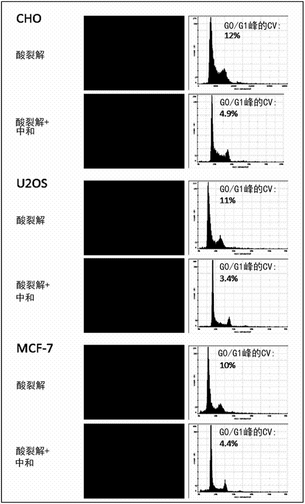
[0312] Example 1. Nuclei Released by Acid Cleavage and Used for Accurate and Precise DNA Quantification

[0313] The DNA content of three adherent cell lines CHO, MCF-7 and U2OS was measured by staining nuclei released by lysis with DAPI (4',6-diamidino-2-phenylindole) acid.

[0314] Materials and methods:

[0315] Adherent cell lines were grown to 80-90% confluency in RPMI+10% FCS in T25 flasks. To release nuclei directly from attached cells, wash the T25 flask once with 5 mL of PBS and then at 37 °C in 0.65 ml of lysis buffer (50 mM citric acid (pH 2.20), 1% Triton X-100, 1 μg / ml DAPI) Incubate in presence for 5 minutes. Fluorescence of released nuclei was either analyzed directly by quantitative imaging cytometry or neutralized by adding an equal volume (0.65 ml) of stabilization buffer (250 mM citrate (pH 8.3), 1% Triton X-100) prior to imaging. and. Quantitative imaging cytometry was performed using a Nucleocounter NC-3000 (Chemometec).

[0316] result:

[0317]...

Embodiment 2

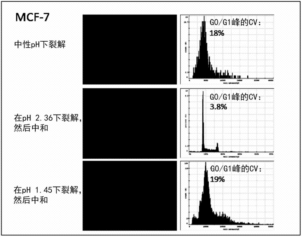
[0321] Example 2, Determination of the Optimal pH of Nuclear Release, Deaggregation and Uniform DNA Staining

[0322] Three adherent cell lines CHO, MCF-7 and U2OS were used to determine the optimum pH for the following three parameters:

[0323] Efficient release of nuclei (>90 nuclei released from attached cells)

[0324] Little or no aggregation of released nuclei

[0325] Uniform DNA staining that provides high accuracy and precision in DNA content measurement

[0326] Materials and methods:

[0327] Adherent cell lines were grown to 80-90% confluency in RPMI+10% FCS in 6-well plates (Nunclon surface, Nunc). To release nuclei directly from attached cells, wash each well once with 1 mL of PBS and dissolve in 0.25 ml of lysis buffer (0.5% Triton X-100, 1 μg / ml DAPI dissolved in 200 mM citric acid (pH 2.00) at 37°C. , 100 mM citrate buffer (pH 2.22-6.80) or dissolved in 400 mM phosphoric acid (pH 1.45)) for 5 minutes. The released nuclei were neutralized by the additio...

Embodiment 3

[0339] The influence of embodiment 3, DAPI concentration and dyeing method

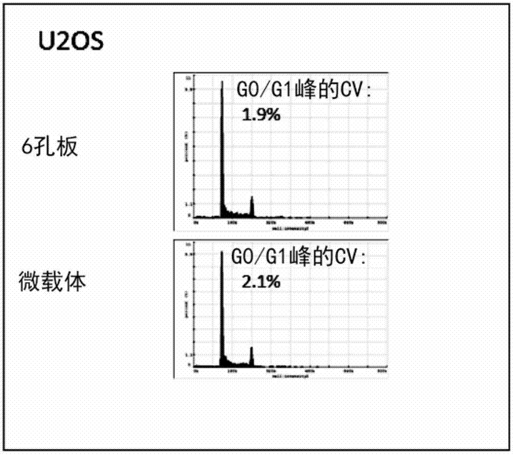
[0340] Whether the accuracy and precision of DNA staining using the adherent cell line U2OS could be further improved by performing nuclear staining in the neutralization step and not in the lysis step.

[0341] Materials and methods:

[0342] U2OS cells were grown to 80-90% confluency in RPMI+10% FCS in 6-well plates. To release nuclei directly from attached cells, wash the wells once with 1 mL of PBS and then in 0.25 ml of lysis buffer (100 mM citrate buffer (pH 2.25), 37 °C, in the presence or absence of DAPI, 0.5% Triton X-100) for 5 minutes. The released nuclei were neutralized by incubation with 0.25 ml stabilization buffer (500 mM citrate, pH 8.3) at 37°C in the presence or absence of DAPI. See Table 2 for details on the experimental conditions, which shows the effect of DAPI concentration and staining method. Fluorescence of nuclei was analyzed by imaging cytometry using a Nucleocounter ...


PUM


Abstract

Disclosed is a fast and simple method for quantification of nucleic acid of biological cells as 2 -step protocol. In the first step cells are treated with an acidic solution containing a non- ionic detergent and a fluorescent DNA specific label. In the second step the sample may be neutralised. Determining of the content of nucleic can be performed by fluorescence microscopy. The method may also be used for obtaining information of cell cycle analysis, ploidy determination, measurements of nucleotide incorporation and assays for proliferation, health, stress level, apoptosis, necrosis, or other state of conditions of cells. The invention also relates to a kit of parts comprising an acidic agent, a detergent, a labelling agent and optionally a neutralization agent.

Description

[0001] All patent and non-patent literature cited in or in this application are hereby incorporated by reference in their entirety. technical field [0002] The present invention relates to a method for determining the amount of nucleic acid in biological cells. The method is simple, fast and reliable to analyze and quantify the DNA or DNA and RNA content of biological cells. The method described here is based on lysing biological cells by adding acid to the sample, labeling the nucleic acids and optionally neutralizing the sample prior to examination of the labeled nucleic acids. This method is especially suitable for use with fluorophores capable of identifying DNA and / or RNA as labeling reagents. The method can be used, for example, in cell cycle analysis, ploidy determination, and analytical assays of the proliferation, health, stress level, apoptosis, necrosis, or other state of cells. The present invention also relates to a device for determining the amount of nucleic ...

Claims


Application Information

Patent Timeline
Patent Type & Authority Applications(China)
IPC IPC(8): G01N1/30C12Q1/68G01N15/14
CPCG01N21/6428C12Q1/6806C12Q1/6895G01N2021/6439C12Q2545/114C12Q2527/125C12Q2527/119
Inventor 索伦·克加拉夫马丁·格伦斯毕尔吉
Owner CHEMOMETEC AS
Why Eureka
Try Eureka