
High-resolution tomography optical microscopic imaging device for tissue samples

A tissue sample, high-resolution technology, applied in sampling devices, measuring devices, material analysis through optical means, etc., can solve problems such as the influence of optical imaging results, interference background, etc.

Inactive Publication Date: 2013-07-17
HUAZHONG UNIV OF SCI & TECH
View PDF1 Cites 8 Cited by
  • Summary
  • Abstract
  • Description
  • Claims
  • Application Information

AI Technical Summary

Problems solved by technology

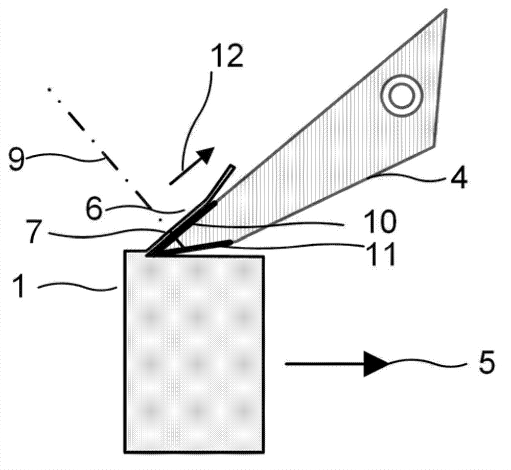
The thin sections of tissue samples cut in this way are spatially separated from the rest of the tissue samples (depending on the thickness of the knife), but since thin sections of plastically embedded tissue samples are usually made of transparent diamond knives, the The remaining sample under the diamond knife will still affect the optical imaging results through the diamond knife, and the interference background will be superimposed on the image

Method used

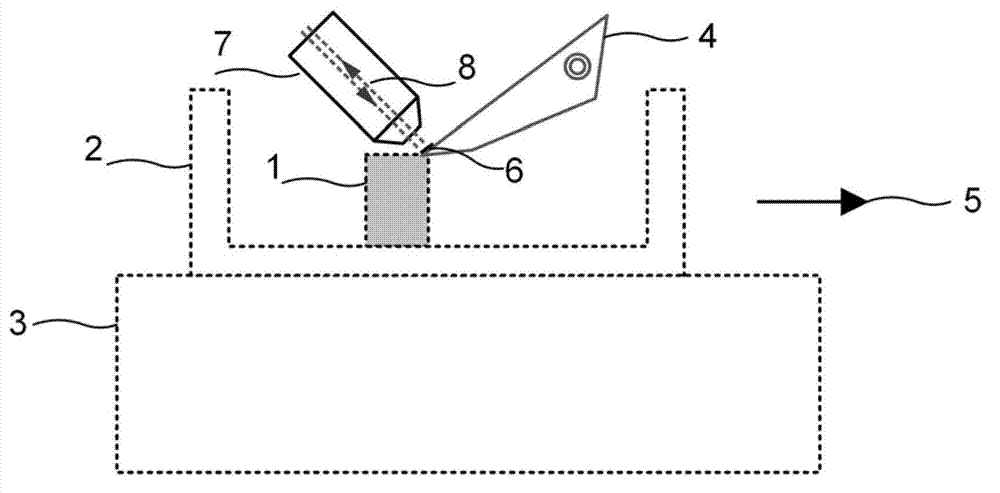
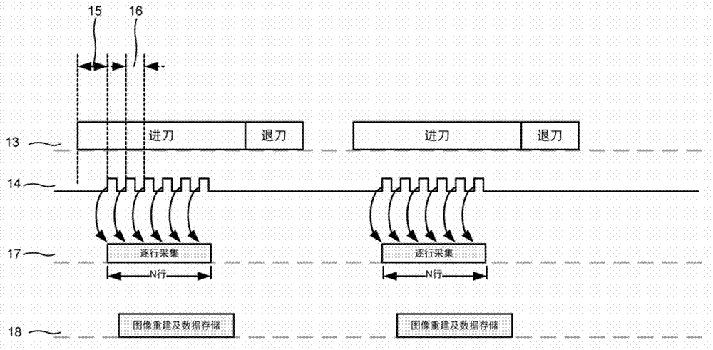

Image

  • High-resolution tomography optical microscopic imaging device for tissue samples
  • High-resolution tomography optical microscopic imaging device for tissue samples
  • High-resolution tomography optical microscopic imaging device for tissue samples

Examples


Embodiment 1

[0026] Such as Figure 4Shown is a mechanically ablated high-resolution tomographic optical microscopy system of a green fluorescent protein (GFP)-labeled tissue sample. The laser 19 outputs laser light with a wavelength of 488nm, passes through the cylindrical lens 20, and focuses the light beam at the focal length of the cylindrical lens 20 into a line light source. An excitation line is formed on the tissue slice 6 through the tube lens 21 and the objective lens 8 . The fluorescence signal of the sample excited by the excitation line is imaged on the photosensitive surface of the line array detector 24 through the objective lens 8 and the tube lens 21 to obtain one row of the fluorescence image of the slice 6 . As the slice moves, the slice 6 slides along the surface of the cutter 4 and is excited line by line by the 488nm excitation line. During the movement of the electric control platform along the direction 5, a trigger signal is generated for each moving distance of t...

Embodiment 2

[0029] The imaging system structure is as in embodiment 1, such as Figure 4 shown. Wherein the cutter 4 uses opaque tungsten carbide hard cutter or black diamond cutter. Because of the opacity of the tool material itself, there is no need to perform optical coating on the tool surface.

[0030] Other settings and steps of the imaging process are the same as those in Embodiment 1, so they are not repeated here.


PUM


Abstract

The invention discloses a high-resolution tomography optical microscopic imaging device for tissue samples. The high-resolution tomography optical microscopic imaging device based on mechanical cutting can cut the tissue samples into thin slices and simultaneously carry out microscopic optical imaging for the thin slices. The device comprises a tissue sample cutting module and an optical microscopic imaging system. A cutting tool of the device is subjected to opacity treatment. The high-resolution tomography optical microscopic imaging device has the advantages that the slices are physically separated from remaining tissue samples in a cutting procedure while tomography images are acquired, illumination light or excitation light of the optical microscopic system cannot be transmitted through the thin slices and the cutting tool to illuminate or excite the remaining tissue samples below the cutting tool owing to the opacity treatment for the cutting tool, and accordingly interference of the tissue samples which are not cut currently on sampling for the slices is eliminated. The high-resolution tomography optical microscopic imaging device can be used for quick high-resolution tomography microscopic imaging for tissue samples.

Description

technical field [0001] The invention is a device for realizing tomographic microscopic imaging of tissue samples based on mechanical cutting. Background technique [0002] High-resolution tomographic optical microscopy imaging of tissue samples, followed by three-dimensional reconstruction, can obtain the three-dimensional spatial distribution of subcellular structures. For neural tissues, the three-dimensional spatial distribution of dendrites and axons of specific neurons can be tracked. To understand the interconnection relationship between neurons. [0003] In order to realize tomographic optical microscopic imaging of tissue samples, optical tomographic imaging technology can be used: confocal imaging technology, through the use of confocal pinholes to block signals outside the focal plane, to achieve tomography; two-photon microscopic imaging technology, using non- Linear effect, the effective optical signal only comes from the focus position to achieve tomography; in...

Claims


Application Information

Patent Timeline
Patent Type & Authority Applications(China)
IPC IPC(8): G01N21/17G01N1/06
Inventor 曾绍群骆清铭龚辉吕晓华熊汗青
Owner HUAZHONG UNIV OF SCI & TECH
Learn More