Liver image segregation method based on hierarchy vessel tree division

A liver segmentation and vascular tree technology, applied in image analysis, image data processing, instruments, etc., can solve the problems of large amount of calculation, parameter adaptive adjustment, influence, etc., to reduce the risk of surgery, strong robustness, The effect of good grading

Inactive Publication Date: 2013-08-28
TONGJI UNIV
View PDF6 Cites 14 Cited by
  • Summary
  • Abstract
  • Description
  • Claims
  • Application Information

AI Technical Summary

Problems solved by technology

Oliveira Dário, Feitosa Raul, Correia Mauro published an article entitled Automatic Couinaud Liver and Veins Segmentation from CT images in BioMedical Engineering OnLine Volume 10, Issue 1 in 2011, and proposed to use the three main branches of the liver portal vein (middle hepatic vein, left hepatic vein) , right hepatic vein) and hepatic portal vein to construct four planes to segment the liver. This method is a typical plane cutting method, but it does not consider the influence of blood vessel variation on the plane selection, and ignores liver segments The fact that the separation between them should be a curved surface; Dirk Selle, Bernhard Preim, Andrea Schenk, Heinz-Otto Peitgen published an article titled Analysis of vasculature for liver surgical planning in Volume 21, Issue 11 of IEEE Transactions on Medical Imaging in 2002, and proposed to adopt The method of calculating the nearest neighbors of different branch vessels to realize liver segmentation. The scale factor parameters between different levels of vessels in this method are manually preset, and the parameter setting cannot be adjusted adaptively for the instance. The accuracy of the grading results, so as to obtain unreasonable segmentation results; Andrea Schenk, Stephan Zidowitz, Holger Bourquain, Milo Hindennach published in volume 6915 of the conference proceedings SPIE Medical Imaging 2008: Computer-Aided Diagnosis entitled Clinical relevance of The article of model based computer-assisted diagnosis and therapy uses the Laplace model to divide the space of blood vessel branches and realize the segmentation of the liver. Relying on the calibration of blood vessel branches affects the robustness of the segmentation method; Wan Shaopeng published a master's thesis entitled Liver Segmentation Method Based on CT Images in 2007, and proposed a three-dimensional visualization based on the skeletonization of the hepatic portal vein As a result, the branches connecting two nodes in the vascular tree are interactively marked to complete the division of vascular branches, and the centerline of various branches is used as the target to calculate the voronoi diagram of the liver space and mask out the segmentation of each liver segment method, this method is essentially a functional segmentation method, the main limitation is that it is time-consuming to interactively mark branches, especially When there are many secondary branches in the vascular tree that need to be marked, it is difficult to meet the real-time requirements, and the branches of the hepatic portal vein cover each other in three-dimensional space, which makes it very difficult to mark the branches; Shao-hui Huang, Bo-liang Wang, Ming Cheng, Wei-li Wu, Xiao-yang Huang, Ying Ju published an article titled A Fast Method to Segment the Liver According to Couinaud's Classification in volume 4987 of the conference proceedings LNCS Medical Imaging and Informatics in 2008, and proposed A fast division method based on projection. This method first constructs a projection plane based on the spatial distribution of the hepatic veins, and divides the projection plane into different mapping regions based on the projection of specific blood vessels on the projection plane to divide the liver. This method is fast and efficient. High, but the idea of ​​functional segmentation is not highlighted while considering the spatial distribution of blood vessels, making the final liver segmentation results not completely consistent with the actual blood supply branches

Method used


Image

  • Liver image segregation method based on hierarchy vessel tree division
  • Liver image segregation method based on hierarchy vessel tree division
  • Liver image segregation method based on hierarchy vessel tree division

Examples


Embodiment 1

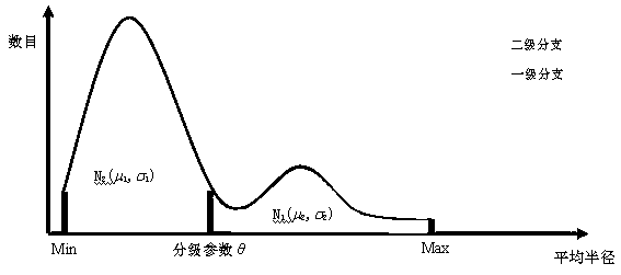
[0018] Example 1: Refer to the attached drawings, in figure 1 A flow chart of the method of the present invention is given in, and a set of embodiments are given according to the illustrated flow. This method first uses a directed tree G=(V, E) to represent the topological structure of the hepatic portal vein vessels, and then according to the spatial distribution of the hepatic portal vein vessels in the specific case images and the average branch radius information, the vessel classification parameters are determined and the hierarchical vessel tree is constructed T 1 And T 2 , Mark the secondary subtree set T that supplies the liver ’ , Divide the marked secondary subtree into eight types according to the blood supply area ’ ={C 0 ,...,C 8 }, and then use the shortest distance algorithm to divide the liver into eight liver segments L={L 0 ,...,L 8 } And perform interpretation and visualization.

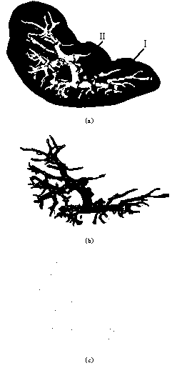
[0019] in figure 2 In the illustrated embodiment, figure 2 (a) is the liver ...


PUM


Abstract

The invention relates to a liver image segregation method based on hierarchy vessel tree division. The method comprises the steps that firstly, a directed tree is utilized to represent a topological structure of a liver portal vein vessel, then according to space distribution and branch average radius information of the liver portal vein vessel in a specific clinical case image, vessel hierarchical parameters are determined and a hierarchical vessel tree is created, a second-level subtree set providing blood for a liver is marked, marked second-level subtrees are divided into eight kinds according to blood supplying areas, the liver is divided into eight hepatic segments through the adoption of the shortest distance algorithm to be analyzed, and clinical interesting information is extracted. According to the method, for liver vessels with multiple branches and complex structures, good hieratical effects can be obtained, blood supplying effects of most second-level branches are considered, and hepatic segment distribution and attribute information obtained through segregation also accord with the Couinaud hepatic segmentation theory.

Description

Technical field [0001] The invention belongs to the field of computer-assisted diagnosis and treatment of liver tumors, and specifically relates to a liver image segmentation method based on hierarchical vascular tree division. Background technique [0002] With the development of liver resection and the advancement of computer-assisted medical technology, the functional segmented virtual anatomy of the liver has attracted more and more attention. Oliveira Dário, Feitosa Raul, Correia Mauro published an article entitled Automatic Couinaud Liver and Veins Segmentation from CT images in BioMedical Engineering OnLine in Vol. , Right hepatic vein) and hepatic portal vein as the basis to construct four planes to segment the liver. This method is a typical plane cutting method, but does not consider the influence of blood vessel variation on the selection of planes, and ignores the liver segment The separation between the two should be the fact that the surface should be a curved surf...

Claims


Application Information

Patent Timeline
Patent Type & Authority Applications(China)
IPC IPC(8): G06T7/00
Inventor 陈宇飞赵卫东王志成柳先辉卫刚岳晓冬
Owner TONGJI UNIV
Why Eureka
Try Eureka