Rapamycin-induced regulatory γδt cell culture method

A technology of rapamycin and a culture method, which is applied in the field of immunology research to achieve the effects of strong feasibility, strong immunosuppressive function, and good promotion value

Active Publication Date: 2016-02-03
ZHEJIANG UNIV
View PDF3 Cites 0 Cited by
  • Summary
  • Abstract
  • Description
  • Claims
  • Application Information

AI Technical Summary

Problems solved by technology

However, currently there are few in vitro induction and expansion methods for γδTreg. Therefore, finding a new in vitro induction and culture method for γδTreg and improving its in vitro induction and expansion efficiency are of great significance to the further research and clinical application of γδTreg in the future.

Method used


Image

  • Rapamycin-induced regulatory γδt cell culture method
  • Rapamycin-induced regulatory γδt cell culture method
  • Rapamycin-induced regulatory γδt cell culture method

Examples


Embodiment 1

[0020] Example 1 Preparation of mononuclear cells derived from human peripheral blood

[0021] 1. Dilute the collected heparin anticoagulated peripheral blood (Zhejiang Blood Center) with the same amount of PBS (phosphate buffered saline) at 1:1;

[0022] 2. Take several 15ml centrifuge tubes, add 5ml of human lymphocyte separation solution to each, and slowly superimpose the diluted peripheral blood on the lymphocyte separation solution along the tube wall. Add 10ml of peripheral blood to each centrifuge tube, that is, 5ml lymph. Cell separation solution + 10ml diluted peripheral blood (1:2); horizontal centrifugation, 500g×20 minutes, room temperature;

[0023] 3. After centrifugation, the tube is divided into three layers, the upper layer is plasma and PBS buffer, the lower layer is mainly red blood cells and granulocytes, the middle layer is lymphocyte separation liquid, and there is a white layer dominated by mononuclear cells at the interface of the upper and middle layers. T...

Embodiment 2

[0026] Example 2 Induction and culture of regulatory γδT cells

[0027] 1. Divide the cultured cells into three major groups, including the IL-2 culture group alone (γδT group), the TGF-β1 / IL-2 / IL-15 induction group (TGF-β1 group), and the combined TGF- β1 / IL-2 / IL-15 and rapamycin induction group (combined with rapamycin group); each cytokine was added on the 0th day of cell culture, and the final concentration after addition is shown in Table 1 below; Rapamycin was also added on day 0 of cell culture.

[0028]

[0029] 2. On the 3rd, 6th, and 9th days, perform a half-volume exchange and add each corresponding cytokine to keep the final concentration of each cytokine consistent.

[0030] 3. The storage concentration of each cytokine are: Zoledronic acid (56mmol / ml), IL-2 (2×10 4 U / ml), IL-15 (10μg / ml), TGF-β1 (1μg / ml), rapamycin (10μM); store at -20°C.

Embodiment 3

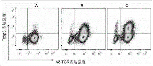
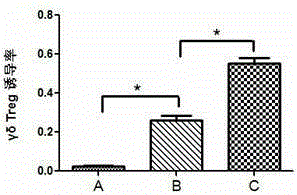
[0031] Example 3 Analysis and comparison of the induction of regulatory γδ T cells under different culture conditions

[0032] At present, the in vitro induction of regulatory γδ T cells is more commonly used by the method of IL-2 / IL-15 / TGF-β1 induction. This experiment is based on IL-2 / IL-15 / TGF-β1 and further combined applications Rapamycin is synergistically induced, and the changes in its induction efficiency are tested and compared.

[0033] 1. Collect cells cultured for 15 days under different induction culture conditions, that is, only the IL-2 culture group (γδT group, as a control), and the TGF-β1 / IL-2 / IL-15 culture group (TGF-β1 group) ) And the combined application of rapamycin and TGF-β1 / IL-2 / IL-15 culture group (combined rapamycin group), flow cytometry detection and analysis of the content of regulatory γδT cells.

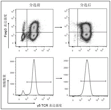
[0034] 2. The method for detecting the content of regulatory γδT cells by flow cytometry is as follows:

[0035] (1) The harvested cells are placed in a fl...


PUM


Abstract

The present invention provides a method for culturing rapamycin-induced regulatory γδT cells, which uses multiple cytokines combined with macrolide immunosuppressant rapamycin to induce human peripheral blood-derived mononuclear cells in vitro Expansion, and a large number of regulatory γδT cells were cultivated. The whole induction, amplification and enrichment process of the present invention is simple and has strong feasibility; the present invention has short cell culture cycle and low cost; high induction efficiency and good repeatability, and can be applied in regulatory γδT cell culture. The regulatory gamma delta T cells induced by the invention have good vigor and strong immunosuppressive function, which can ensure further research in the future and have good popularization value.

Description

technical field [0001] The invention belongs to the field of immunology research, and relates to a method for culturing regulatory γδT cells induced by rapamycin, which is to apply various cytokines (TGF-β1 / IL-15 / IL-2) combined with macrolides to immunize The inhibitory drug, rapamycin, synergistically induces the generation and expansion of regulatory γδ T cells. Background technique [0002] γδT cells are a special subpopulation of T lymphocytes, which have unique structures and biological functions. Different from general αβT cells, γδT cell surface receptors are composed of two glycoprotein chains, γ chain and δ chain, hence the name; this group of cells does not need to be presented by histocompatibility antigen molecules on the cell surface to recognize antigens. Direct recognition of protein, peptide and non-peptide antigens, that is, the process is non-HLA (human leukocyte antigen) restricted, is an important biological characteristic of γδT cells. Although the pro...

Claims


Application Information

Patent Timeline
Patent Type & Authority Patents(China)
IPC IPC(8): C12N5/0783
Inventor 黄河顾嫣珺胡永仙
Owner ZHEJIANG UNIV
Why Eureka
Try Eureka