Compositions and methods to prevent cell transformation and cancer metastasis

A cancer, transferase technology, applied in the field of cancer diagnosis and treatment, can solve the problems of poorly understood and unsatisfied importance

Inactive Publication Date: 2013-12-25
CORNELL UNIVERSITY
View PDF2 Cites 0 Cited by
  • Summary
  • Abstract
  • Description
  • Claims
  • Application Information

AI Technical Summary

Problems solved by technology

However, how these structures arise and their importance in cancer progression is currently poorly understood
Therefore, there is a continuing and unmet need to explore the heretofore unrecognized roles of MVs in cancer and to exploit these roles to develop compositions and methods involving diagnostic methods and therapeutic interventions

Method used


Image

  • Compositions and methods to prevent cell transformation and cancer metastasis
  • Compositions and methods to prevent cell transformation and cancer metastasis
  • Compositions and methods to prevent cell transformation and cancer metastasis

Examples


Embodiment 1

[0116] The results in this example were obtained from experiments using the materials and methods described in Example 3.

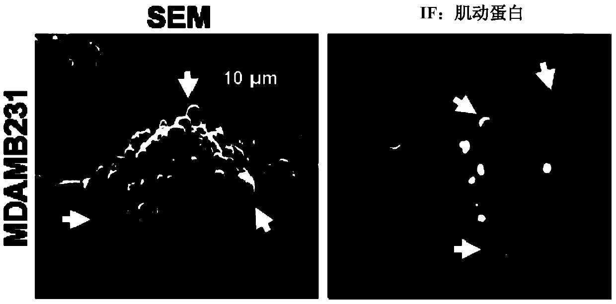
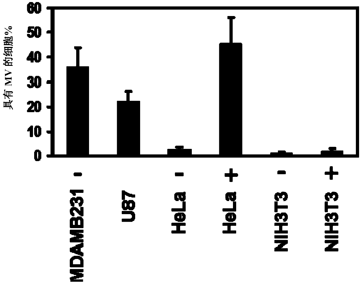
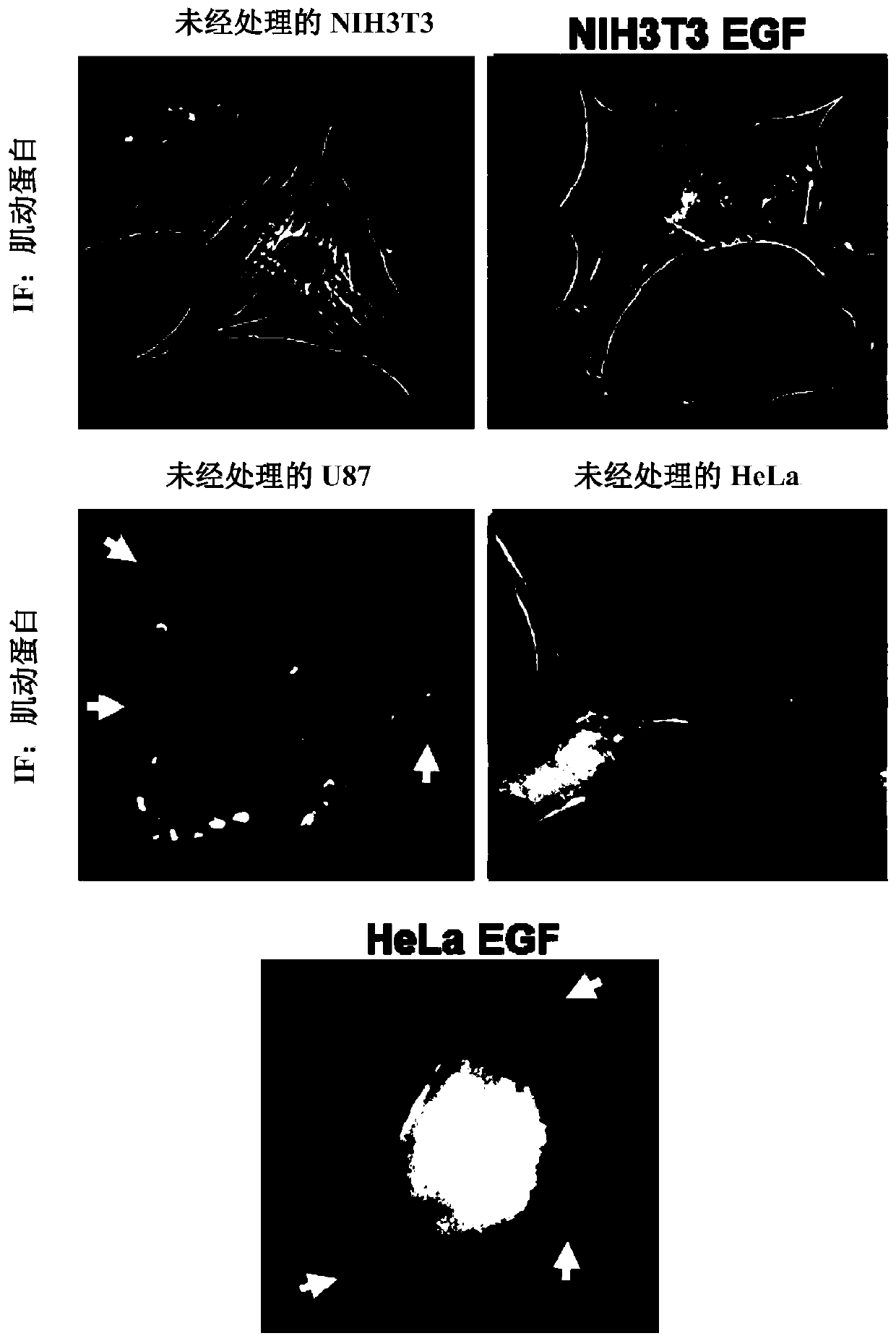
[0117] We used scanning electron microscopy (SEM) ( figure 1 , left panels) or using fluorescence microscopy on cells stained for F-actin ( figure 1 , right) Analysis of serum-starved cultures of the highly invasive human breast cancer cell line MDAMB231 revealed ~35% of the cell surface to have MVs ranging in size from ~0.2-2.0 microns in diameter ( figure 2 ). MVs were also detected on ~25% of serum-depleted U87 human glioma cells, and the formation of these MVs needs to be induced by stimulation with epidermal growth factor (EGF) secreted by HeLa cervical cancer cells ( figure 2 and 3 ). In contrast, MVs were not detected on the surface of normal NIH3T3 fibroblasts cultured under serum-starved or EGF-stimulated conditions, suggesting that some cell types may not produce MVs. Moreover, experiments have shown that MVs are actively shed from these ...

Embodiment 2

[0131] This example describes proteins that co-occur in MVs derived from MDAMB231 cells and U87 cells. Proteomic analysis was performed using MVs shed from MDAMB231 breast cancer cells or U87 brain tumor cells. The following list was compiled from those proteins identified in MVs from MDAMB231 and U87 cells (based on general cellular function): Proteomic analysis of microvesicles shed from MDAMB231 and U87 cells: nucleic acid binding proteins; Nuclear translation elongation factor 1; Eukaryotic translation elongation factor 2; Histone cluster 1; Histone cluster 2; RuvB-like protein 1; RuvB-like protein 2; Extracellular matrix and plasma membrane-associated proteins; Annexin A2; CD9 Antigen; collagen; ecto-5'-nucleotidase; protein 3 containing EGF-like repeats and discoid I-like domains; fibronectin; galectin 3-binding protein; integrin β1; laminin protein; lysyl hydroxylase precursor; major histocompatibility complex; Na+ / K+-ATPase; transglutaminase 2 subtype a; metabolic pro...

Embodiment 3

[0133] This example provides a description of the materials and methods used to obtain the results described in Example 1.

[0134] Material. 4,6-diamidino-2-phenylindole (DAPI), brefeldin A, mitomycin-C, and Exo1 were from Calbiochem, and T101 was from Zedira. Rhodamine-conjugated Phalloidin, EGF, Lipofectamine, Lipofectamine2000, Protein G beads, control and tTG siRNA, and all cell culture reagents were from Invitrogen. FN antibody, MDC, and BPA were from Sigma. tTG and actin antibodies were from Lab Vision / Thermo. Lipid raft marker protein-2 antibody was from Santa Cruz, and HA and Myc antibodies were from Covance. Steriflip PVDF-filters (0.45 μm pore size) were from Millipore. Antibodies against IκBα, GFP, and antibodies recognizing ERK, AKT, FAK, and EGF receptors were from Cell Signaling.

[0135] cell culture. MDAMB231, U87, MCF10A, and HeLa cell lines were grown in RPMI1640 medium containing 10% fetal bovine serum, while NIH3T3 cell lines were grown in DMEM mediu...


PUM

PropertyMeasurementUnit
diameteraaaaaaaaaa
diameteraaaaaaaaaa

Abstract

Provided are methods for characterizing microvesicles or other membranous structures. The methods involve assaying samples for microvesicles or other membranous structures, and include in certain aspects determining the presence or absence of tissue transglutaminase (tTG) and / or cross-linked fibronection (FN). The microvesicles or other membranous structures can be separated from a sample using recombinant tTG or a derivative of it, or tTG or FN binding partners. Also provided are methods for inhibiting the transfer of cargo from microvesicles which contain tTG to one or more cells. This involves administering to the individual a tTG inhibitor, such as a cell-impermeable tTG inhibitor. Also provided are compositions which contain a population of microvesicles or other membranous structures, where the population is attached to tTG or a derivative thereof, or to tTG or an FN binding partner. Kits which contain reagents and other components for carrying out the methods are also provided.

Description

[0001] Cross References to Related Applications [0002] This application claims priority to US Provisional Patent Application No. 61 / 443,978, filed February 17, 2011, the disclosure of which is incorporated herein by reference. field of invention [0003] The present invention relates generally to the diagnosis and treatment of cancer, and more particularly to novel cancer markers and therapies based on microvesicles shed from cancer cells. Background of the invention [0004] Tumor progression involves the ability of cancer cells to communicate with each other and with neighboring normal cells in their microenvironment. Microvesicles (MVs) derived from human cancer cells have attracted attention because of their apparent ability to participate in the horizontal transfer of signaling proteins between cancer cells and to promote the invasive activity of cancer cells. Different types of advanced or invasive forms of human cancer cells release MVs into their surrounding envir...

Claims


Application Information

Patent Timeline
Patent Type & Authority Applications(China)
IPC IPC(8): G01N33/53
CPCG01N33/5076G01N33/57484G01N33/6893
Inventor R·塞立昂M·安东雅克W·K·塞立昂李波
Owner CORNELL UNIVERSITY
Why Eureka
Try Eureka