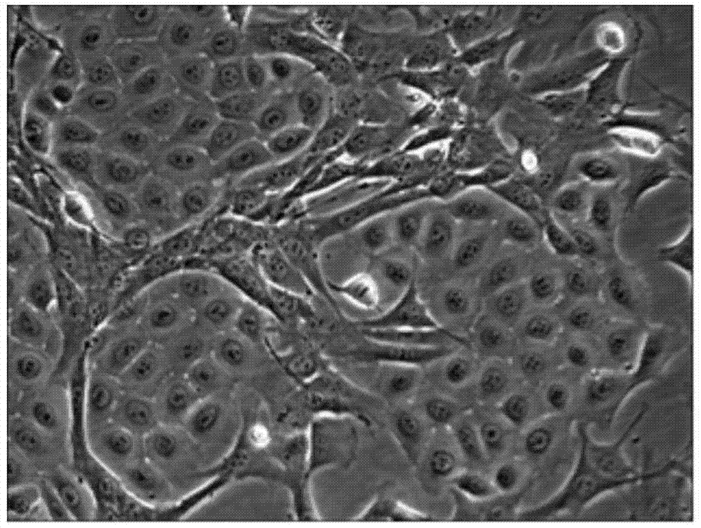
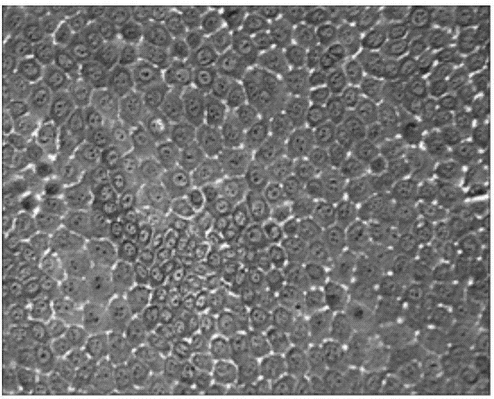
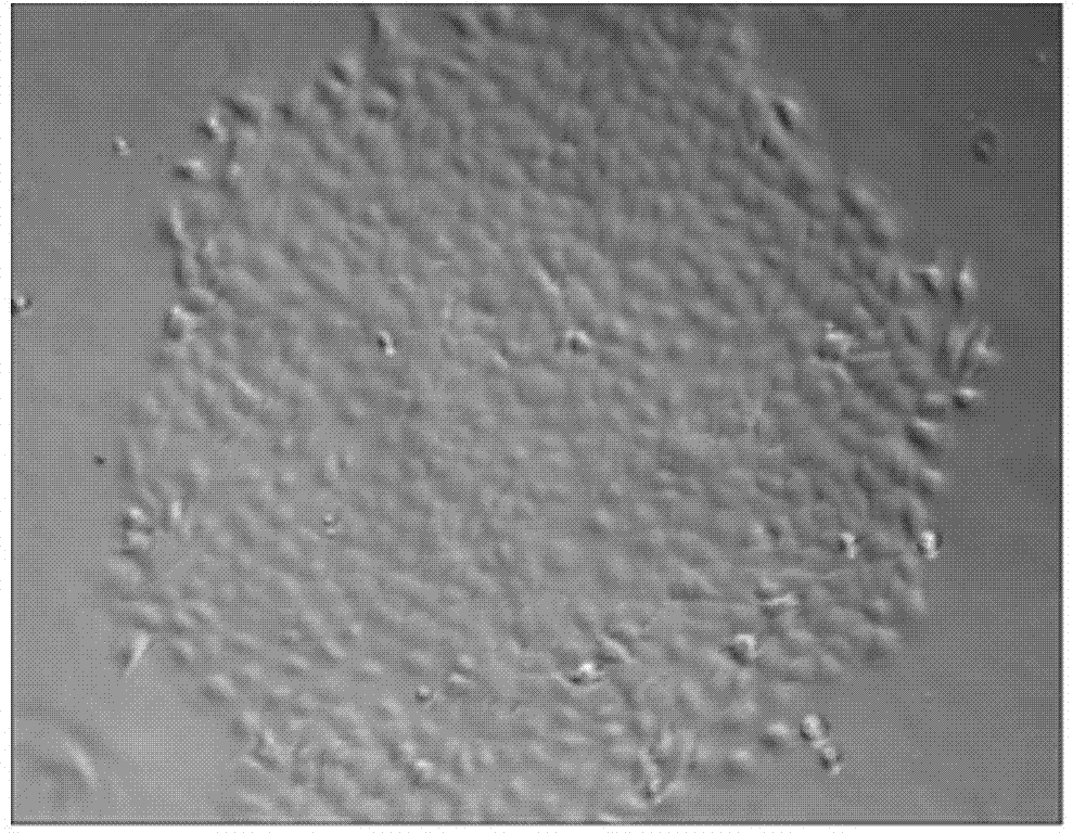
Single cell cloning method for obtaining goat mammary epithetical cells
A single-cell cloning, goat mammary gland technology, applied in animal cells, vertebrate cells, artificial cell constructs, etc., can solve the problem of low cell clone formation rate, and achieve the effect of improving the single-cell clone formation rate
- Summary
- Abstract
- Description
- Claims
- Application Information
AI Technical Summary
Problems solved by technology
Method used
Image
Examples
Embodiment 1
[0059] Example 1 Sampling and Processing of Goat Mammary Epithelial Cells
[0060] The mammary gland tissues of the purebred Laoshan dairy goats of Qingdao Aotai sheep farm in the lactation period of 30 days were directly aseptically collected by using the existing technology;
[0061] Rinse the collected breast tissue block with PBS to remove the blood and goat milk on its surface; after rinsing, immerse it in DF12 medium containing 200U / mL penicillin and 200μg / mL streptomycin, put it in an ice box and quickly bring it back to the laboratory For treatment, the breast tissue was washed repeatedly with PBS containing 200 U / mL penicillin and 200 μg / mL streptomycin until the washing liquid became clear, divided and trimmed on an ultra-clean bench, and the adipose tissue and connective tissue were removed and rinsed until the washing liquid was clear. White granular acinar tissue can be obtained.
Embodiment 2
[0062] Example 2 Tissue Block Culture Isolation of Mammary Epithelial Cells and Fibroblasts
[0063] a. Use ophthalmic scissors and ophthalmic forceps to divide the above-mentioned white granular acinar tissue into 0.5-1mm 3 The left and right small tissue pieces were rinsed with PBS containing 200 U / mL penicillin and 200 μg / mL streptomycin;
[0064] b. Infiltrate the small tissue pieces with the medium prepared by FBS and DF12 at a volume ratio of 1:1 for 2-4 minutes, inoculate them on the bottom of the culture dish at an interval of 0.5 cm, and place them at 37°C and 5vt% CO 2 , in an incubator with saturated humidity for 4 hours;
[0065] c. Add culture medium to the above-mentioned petri dish so that it just covers the bottom of the petri dish, and place at 37°C, 5vt% concentration of CO 2 , cultured in an incubator with saturated humidity, add culture solution after 12 hours until the tissue block is completely submerged, replace half of the culture solution after 2 day...
Embodiment 3
[0069] Fibroblasts were removed by rapid digestion combined with differential attachment method to obtain purified mammary epithelial cells. The specific steps are as follows:
[0070] a. Differential digestion method: add digestive fluid to the culture of breast epithelial cells grown with other cells to digest the cells, observe the changes in cell morphology under an inverted microscope, and when the fibroblasts are detached from the bottom wall of the culture dish, aspirate and digest Remove the fibroblasts, use the remaining digestion solution to continue digestion for 1-2 minutes, add culture medium to stop the digestion, and pipette the remaining cells into a single-cell suspension;
[0071] Wherein the digestion solution is an equal volume of PBS without calcium and magnesium ions and 0.25% Trypsin-EDTA solution, the digestion condition is room temperature 25°C, and the digestion time of fibroblasts is 1.5-2 minutes;
[0072] The culture solution is a solution with a v...
PUM
 Abstract
Description
Claims
Application Information
 - R&D
- Intellectual Property
- Life Sciences
- Materials
- Tech Scout
- Unparalleled Data Quality
- Higher Quality Content
- 60% Fewer Hallucinations
Browse by: Latest US Patents, China's latest patents, Technical Efficacy Thesaurus, Application Domain, Technology Topic, Popular Technical Reports.
© 2025 PatSnap. All rights reserved.Legal|Privacy policy|Modern Slavery Act Transparency Statement|Sitemap|About US| Contact US: help@patsnap.com


