
Tumor site identification device and method

A technology for identifying devices and tumors, applied to measurement devices, analysis using fluorescence emission, sensors, etc., can solve problems such as hindering PpIX fluorescence detection, achieve accurate and rapid diagnosis, improve surgical results, and reduce the effect of surgery

Inactive Publication Date: 2014-02-26
KYOTO PREFECTURAL PUBLIC UNIV CORP
View PDF6 Cites 21 Cited by
  • Summary
  • Abstract
  • Description
  • Claims
  • Application Information

AI Technical Summary

Problems solved by technology

[0006] However, in the human body, lymph nodes exist surrounded by connective tissues, and these connective tissues (fat, collagen, etc.) emit strong autofluorescence in the blue to green wavelength region under blue excitation light, which hinders the fluorescence of PpIX detection

Method used


Image

  • Tumor site identification device and method
  • Tumor site identification device and method
  • Tumor site identification device and method

Examples


Embodiment 1

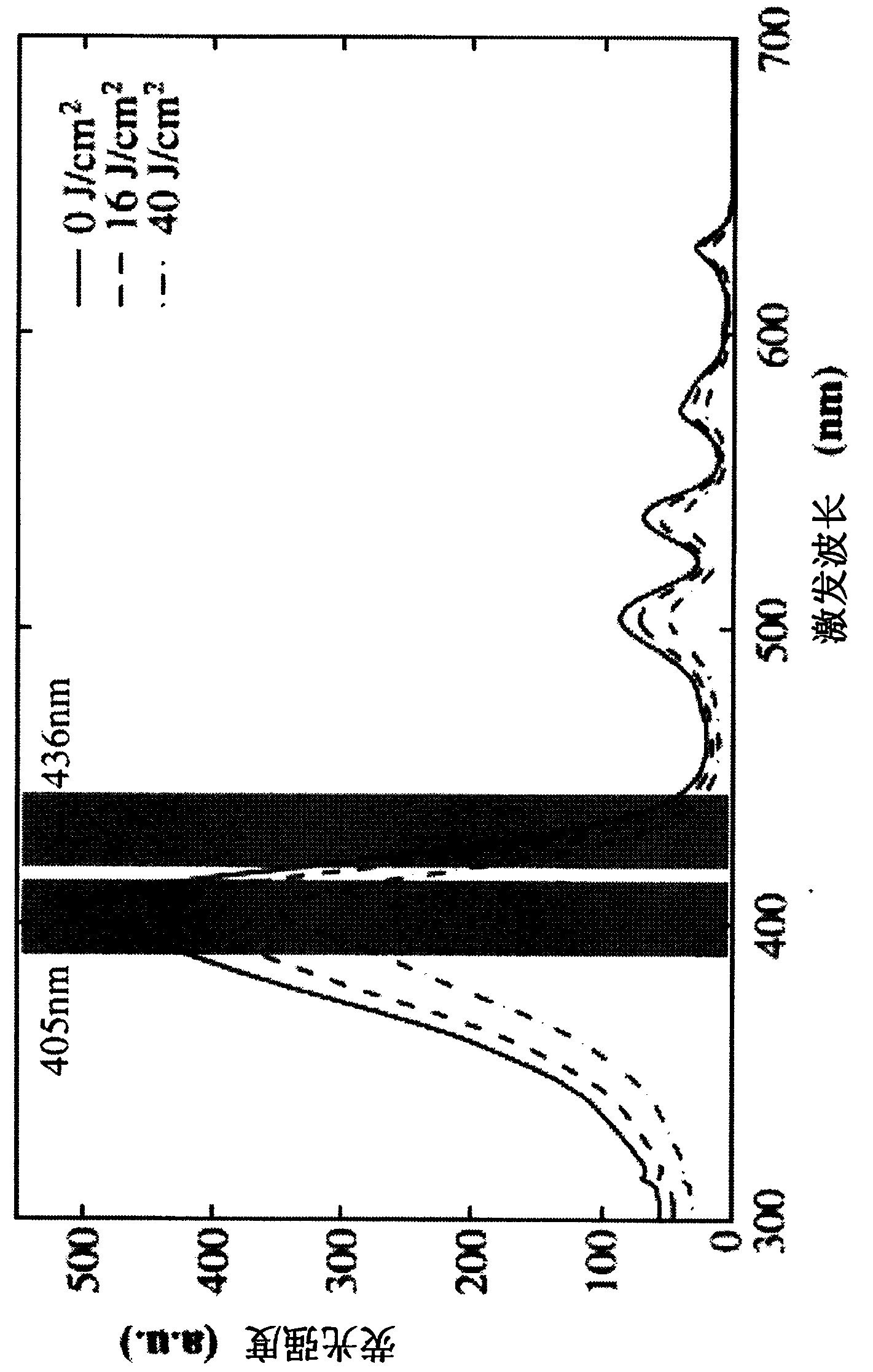
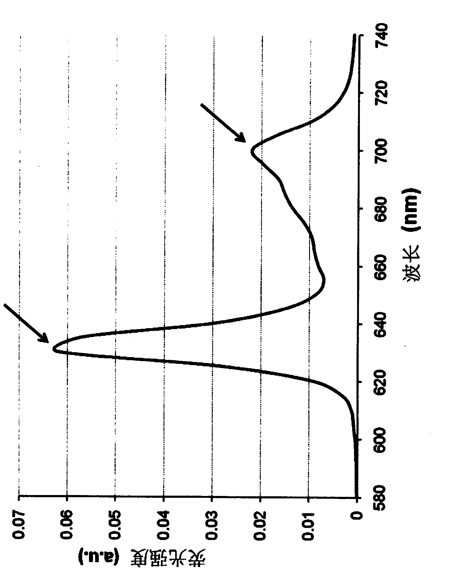
[0133] After administering 5-ALA to the gastric cancer cell line (MKN-45) and culturing for 3 hours, the excitation light of 436 nm was continuously irradiated, and the spectrum was measured at regular intervals. Compared with the PpIX solution, although the rate of change is different, the same tendency (decrease of the peak at 635nm and increase of the peak at 675nm) was observed in cultured cells ( Figure 10 ). In addition, it was confirmed that the ratio (I 675 / I 635 ) depends on the exposure and rises ( Figure 11 ). However, the rate of change was lower than in the case of using the PpIX solution.

Embodiment 2

[0135] To graphically capture spectral changes, ratiometric imaging was performed. That is, under 436nm excitation light, spectral images of 635nm and 675nm were acquired at regular intervals of light irradiation, their ratio images were created, and changes in the ratio images on the left and right sides of irradiation were observed. The result is as Figure 12 shown. Similar to the results of the spectrometry, it was shown that the brightness of the ratio image gradually increased with light irradiation.

Embodiment 3

[0137] In the same manner as in Example 2, ratiometric imaging was performed by simultaneously observing the cell suspension of cancer cells and collagen fibers after administration of 5-ALA.

[0138] Although cancer cells and collagen cannot be distinguished in each spectroscopic image, if each ratio image (I R pre and I R post ), and then make its ratio (I R post / I R pre ), it is clear that only the site where PpIX exists is imaged as a higher value than the surrounding area ( Figure 13 ).


PUM


Abstract

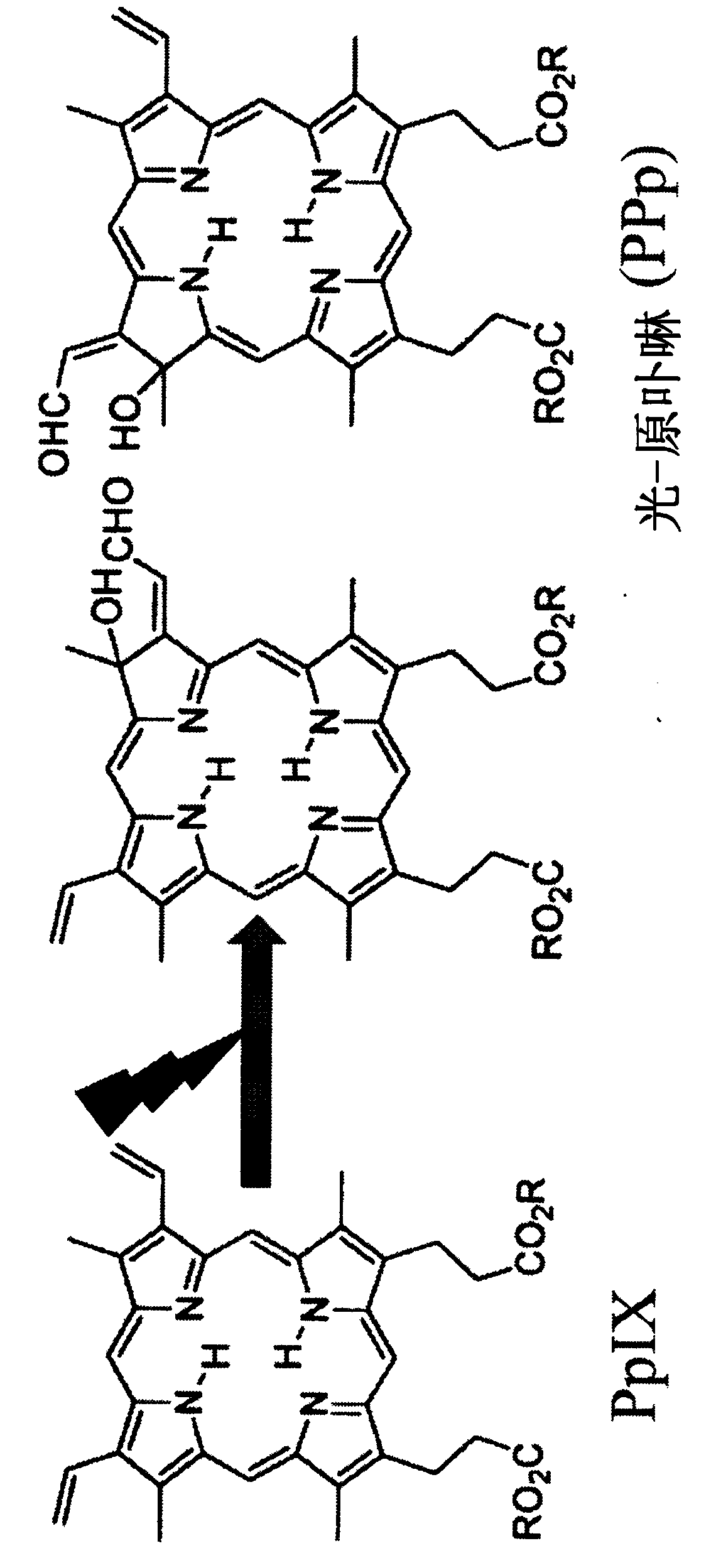
The invention provides a tumor site identification device which spectroscopically detects the fluorescence of protoporphyrins present at a tumor site in a test subject, wherein the protoporphyrins are protoporphyrin IX (PpIX) and photo-protoporphyrin (PPp), and the identification device comprises a light irradiation unit that converts part of PpIX to PPp, a light splitting unit that splits the fluorescence of PpIX and the fluorescence of PPp, a spectroscopic detection unit that detects the relative fluorescence intensities of the fluorescence of PpIX and the fluorescence of PPp, and a tumor discrimination unit that discriminates between a tumor site and a non-tumor site on the basis of the relative fluorescence intensities of PpIX and PPp.

Description

technical field [0001] The invention relates to a tumor site identification device and identification method. Background technique [0002] Lymph node metastasis is one of the important prognostic factors in digestive organ cancers such as gastric cancer and colorectal cancer, and it is very important to accurately diagnose the presence or absence of lymph node metastasis when deciding on the treatment of a patient. [0003] For example, in most digestive organ cancers, the need for postoperative adjuvant therapy is determined based on the presence or absence of lymph node metastasis (Non-Patent Documents 1 and 2). In addition, especially for gastric cancer, the sentinel lymph node biopsy When it is possible to diagnose no metastasis in the examination, surgery to reduce the cleared area may be selected (Non-Patent Document 3). For such reasons, it would be very useful clinically if lymph node metastasis could be diagnosed promptly and accurately. [0004] However, a new d...

Claims


Application Information

Patent Timeline
Patent Type & Authority Applications(China)
IPC IPC(8): G01N21/64
CPCA61B5/0071G01N2021/6421A61B5/00G01N21/6456G01N21/6428G01N21/6458G01N21/6486A61B1/00186A61B1/043A61B1/0646A61B5/0084A61B5/0091A61B5/418A61B5/6847
Inventor 小泉范明高松哲郎原田义规大辻英吾
Owner KYOTO PREFECTURAL PUBLIC UNIV CORP
Learn More