Method and medicament for inhibiting generation of neonatal lymphatic vessel
An inhibitor, selected technology, applied in the direction of drug combination, chemical equipment and methods, antipyretics, etc., can solve ambiguous problems
- Summary
- Abstract
- Description
- Claims
- Application Information
AI Technical Summary
Problems solved by technology
Method used
Image
Examples
Embodiment 1
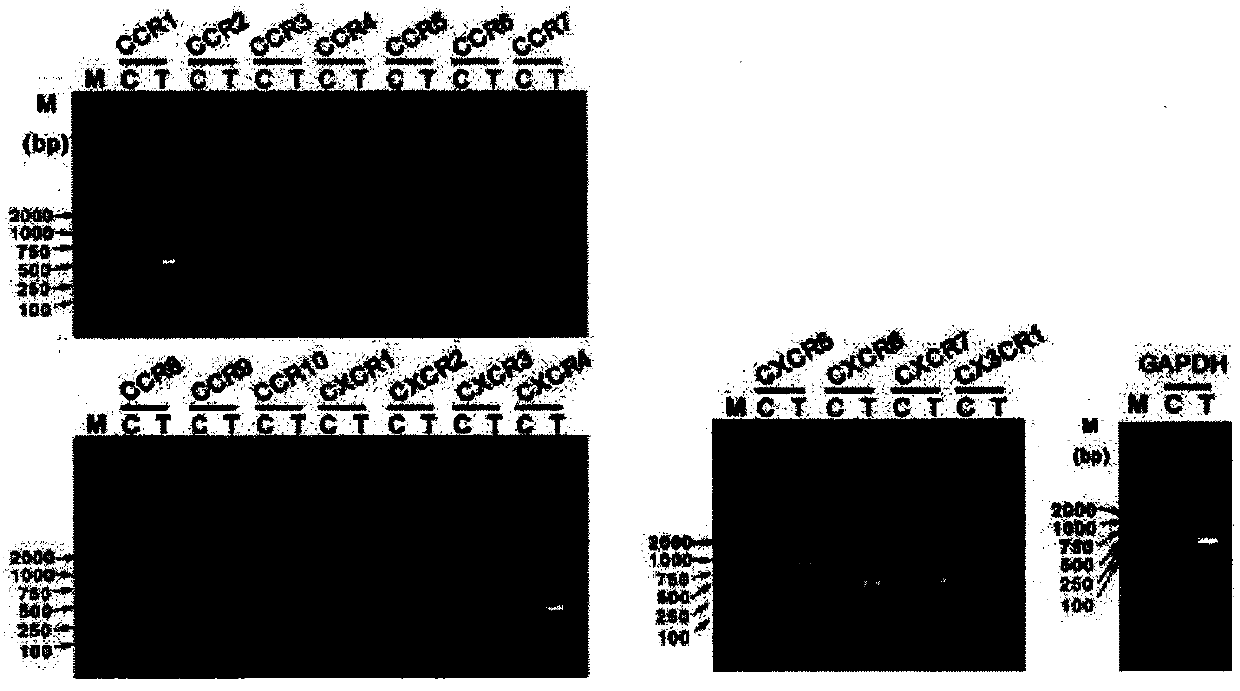
[0072] Lymphatic endothelial cells express multiple chemokine receptors
[0073] experimental method
[0074] 1. Total RNA extraction and RT-PCR detection of chemokine receptor expression on lymphatic endothelial cells
[0075] The isolation and extraction of total cellular RNA was performed using TRIZOL reagent (purchased from Invitrogen) and performed according to the standard operation of the reagent manual. Primary lymphatic endothelial cells collected by centrifugation (approximately 1×10 6 ) into 1mL TRIZOL, blow and suck repeatedly 30 times with the tip of the pipette, and let stand at room temperature for 5 minutes. Centrifuge at 10,000 g at 4°C for 10 minutes, and gently aspirate the supernatant. Add 0.2 mL of chloroform, shake vigorously for about 15 seconds, and let stand at room temperature for 3 minutes. Centrifuge at 10,000g at 4°C for 15 minutes, and the sample is divided into three layers: a yellow organic phase, an intermediate layer, and a colorless aque...
Embodiment 2
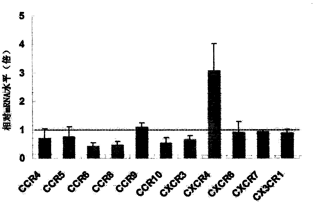
[0119] Chemokine receptor CXCR4 is highly expressed specifically on VEGF-C activated lymphatic endothelial cells
[0120] experimental method
[0121] 1. RT-PCR detection of chemokine receptor expression on lymphatic endothelial cells
[0122] Fluorescence quantitative Real-Time PCR using Stratagene kit (Brilliant II QRT-PCR Master Mix), the fluorescent quantitative PCR instrument is MX3000P (purchased from Stratagene), the fluorescent dye is SYBR Green, the reaction system is 20 μL, and the number of reaction cycles is 40. The internal reference is GAPDH, and the ΔCt value is obtained according to the fluorescence image given by the instrument, and the relative Δ(ΔCt) value is calculated to calculate the relative change of the corresponding gene level.
[0123] 2. Flow cytometry verification of the expression of chemokine receptor CXCR4 on the surface of lymphatic endothelial cells induced by vascular endothelial growth factor C (VEGF-C)
[0124] The mouse primary lympha...
Embodiment 3
[0142] CXCR4 specifically and highly expressed on the surface of neonatal lymphatic vessels in vivo
[0143] experimental method
[0144] 1. Detection of the distribution of chemokine CXCR4 in lymphatic vessels in vivo by tissue immunofluorescence
[0145] Take 8 healthy C57BL / 6 mice (6-8 weeks old, female, purchased from Beijing Weitong Lihua Co., Ltd.), and divide them into two groups. Inoculate 5×10 intradermally 6 Five tumor-bearing mice of B16 / F10 mouse melanoma cells (American Type Culture Collection, ATCC). After 14 days of inoculation, the tumor tissues and paratumor axillary lymph node tissues of tumor-bearing mice, as well as the colon and lymph node tissues of normal mice were removed.
[0146] Fixed embedding. Fix the removed tissue with 4% formaldehyde solution overnight, and then wash the tissue with tap water overnight to remove formaldehyde (you can wrap the tissue block with gauze, put it in a beaker, and wash it overnight with dripping water under the ta...
PUM
 Abstract
Description
Claims
Application Information
 - R&D
- Intellectual Property
- Life Sciences
- Materials
- Tech Scout
- Unparalleled Data Quality
- Higher Quality Content
- 60% Fewer Hallucinations
Browse by: Latest US Patents, China's latest patents, Technical Efficacy Thesaurus, Application Domain, Technology Topic, Popular Technical Reports.
© 2025 PatSnap. All rights reserved.Legal|Privacy policy|Modern Slavery Act Transparency Statement|Sitemap|About US| Contact US: help@patsnap.com


