Organ-type artificial skin as well as preparation method and application thereof

A technology of artificial skin and organs, applied in skin transplantation, medical science, prosthesis, etc., can solve problems such as limited self-renewal ability, cell cycle entering terminal differentiation, and difficulty in exerting synergistic effects of various cells to achieve good cell active effect

Inactive Publication Date: 2014-07-02
BIOPHARM RES & DEV CENT JINAN
View PDF4 Cites 9 Cited by
  • Summary
  • Abstract
  • Description
  • Claims
  • Application Information

AI Technical Summary

Problems solved by technology

However, most of the existing artificial skin models only have two types of cells: keratinocytes and fibroblasts, and there are few studies on models that introduce other skin cells. Due to the limited types of cells, it is difficult to exert the syner...

Method used


Image

  • Organ-type artificial skin as well as preparation method and application thereof
  • Organ-type artificial skin as well as preparation method and application thereof
  • Organ-type artificial skin as well as preparation method and application thereof

Examples


Embodiment 1

[0045] Example 1 Construction of artificial skin

[0046] 1. Materials

[0047] Keratinocytes and fibroblasts were obtained from normal skin after circumcision of children, and amnion was obtained from the placenta of healthy parturients, and frozen after decellularization for future use. BD Matrigel TM BasementMembrane Matrix was obtained from BD Biosciences and human placental collagen type IV was obtained from Sigma.

[0048] 2. Method

[0049] 1) Culture method of human dermal fibroblasts

[0050] After the epidermis was torn off from the dermis, the dermis was washed 3-5 times with PBS containing antibiotics. Use ophthalmic scissors to cut the dermal tissue into a paste, and use ophthalmic forceps to take about 0.5mm of it 3 Tissues of large size were inoculated in a 10cm culture dish, and the interval between each tissue block was about 10mm. The dish was placed upside down in a 37°C incubator for 1 hour. After the tissue block was firmly attached to the bottom of ...

Embodiment 2

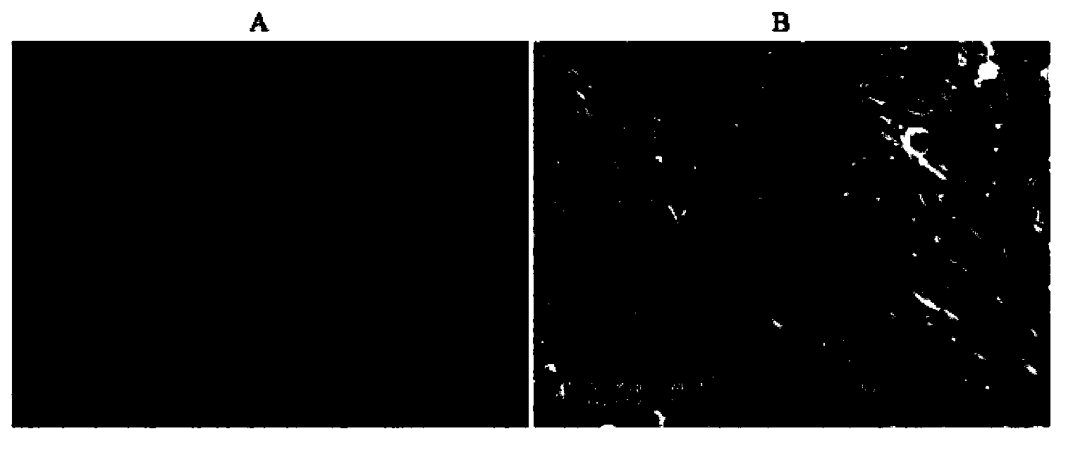
[0056] Example 2 Expression of normal skin-specific proteins in the organotypic artificial skin of the present invention

[0057] As a result of immunohistochemical testing, it was found that the organotypic artificial skin prepared in Example 1 expressed the corresponding protein markers of normal human skin, including involucrin (mantle protein) in the stratum corneum, CK10 in the spinous layer, CK14 in the basal layer, and vimentin in the dermis ( Vimentin) and CK19 of basal epidermal stem cells ( Figure 4A -E). Among them, the stratification of the epidermis and dermis is clear, the distribution of proteins in each layer is basically the same, and the epidermal stem cells are located in the position of the epidermis close to the basal layer. The positive cells of mantle protein and CK10 were less than those of normal skin, suggesting that this program can be further optimized to achieve better results.

Embodiment 3

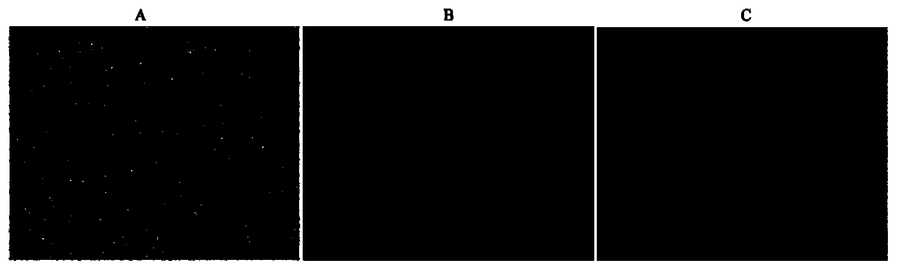
[0058] The cell viability assay of embodiment 3 skin

[0059] The results of trypan blue staining to detect cell viability show that the fibroblasts and keratinocytes grown in the organotypic artificial skin prepared in Example 1 can maintain good cell viability, and the number of viable cells reaches 98% ( Figure 5 ).


PUM

PropertyMeasurementUnit
Thicknessaaaaaaaaaa

Abstract

The invention relates to organ-type artificial skin and a preparation method thereof. The organ-type artificial skin disclosed by the invention is formed by constructing fibroblasts, a human acellular amniotic membrane or keratinocytes or by constructing fibroblasts, amniotic fluid stem cells, a host material and keratinocytes. The artificial skin disclosed by the invention has dermis and epidermis structures close to a normal skin structure and a protein marker expressing normal skin, and distribution of a cuticle, a stratum spinosum, a basal layer and dermis is consistent with that of normal skin; the artificial skin can serve as a skin substitute, skin temporary covering and an artificial skin model.

Description

technical field [0001] The invention belongs to the technical field of artificial organ tissue engineering, and in particular relates to a novel organ-type artificial skin and a construction method thereof. Background technique [0002] In recent years, the construction of organotypic artificial skin with a bilayer structure of epidermis and dermis has been the focus of research. The artificial skin constructed in vitro requires the structure and function to be similar to normal skin, otherwise it is just a layer of cells, which cannot meet the needs of actual research and application. Such as SKIN2 developed by Advanced TissueSciences in the United States TM For the skin, the fibroblasts are planted in the nylon mesh to form the dermis, and the keratinocytes are planted on the dermis to form the epidermis. However, most of the existing artificial skin models only have two types of cells, keratinocytes and fibroblasts, and there are few studies on models that introduce oth...

Claims


Application Information

Patent Timeline
IPC IPC(8): A61L27/60A61L27/22A61F2/10
Inventor 黄亚东张齐好项琪苏志坚戴桃李
Owner BIOPHARM RES & DEV CENT JINAN
Why Eureka
Try Eureka