TfR (transferrin receptor) specific binding peptide and application thereof
A transferrin-binding peptide technology, applied in the direction of peptides, drug combinations, non-active ingredients of polymer compounds, etc., can solve problems such as limitations
- Summary
- Abstract
- Description
- Claims
- Application Information
AI Technical Summary
Problems solved by technology
Method used
Image
Examples
Embodiment 1
[0028] Through the method of phage display, the transferrin receptor TfR specific binding peptide CPU-312-03 was obtained:
[0029] (1) The nucleotide sequence of the transferrin receptor TfR-specific binding peptide CPU-312-03 was obtained by screening the phage random peptide library:
[0030]A. Biopanning of phage random 7-peptide library: Dilute transferrin receptor TfR to 100 μg / ml with coating solution (0.1M NaHCO3, pH8.6), add 150ul to a well of the microplate, repeat Spin until surface is completely wet, leave wet overnight at 4°C. Pour off the coating solution, clap the plate to remove the residual solution, fill up with blocking solution (0.1M NaHCO3, pH8.6, 5mg / ml BSA), and act at 4°C for at least one hour. Pour out the blocking solution, wash 6 times with 0.1% TBST [TBS+0.1% (v / v) Tween-20], and tap the plate each time. Dilute 10 μl of the original phage peptide library with 100 μl TBST, add it to the wells coated with the transferrin receptor TfR, incubate at ro...
Embodiment 2
[0038] In silico simulation of transferrin receptor TfR binding to binding peptide:
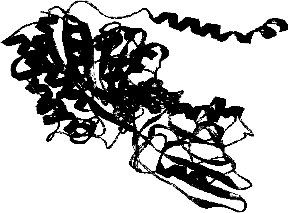
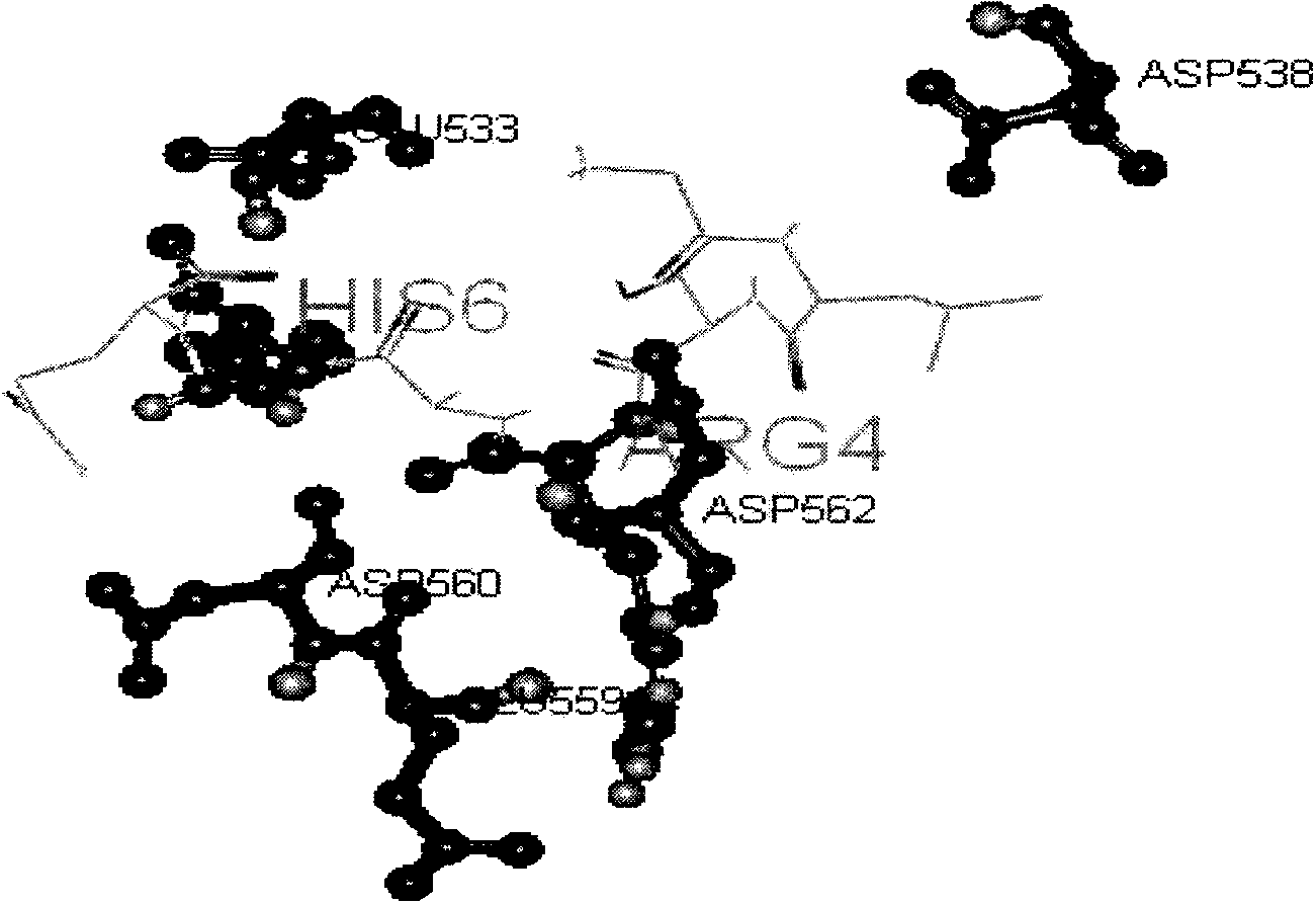
[0039] The model of transferrin receptor TfR and binding peptide was established by zDock docking software, and then the binding situation of transferrin receptor TfR and binding peptide was simulated by discovery studio2.5 (DS2.5) software, transferrin receptor TfR The complex model formed with the binding peptide as figure 1 , where the bars represent the transferrin receptor TfR and the spheres represent the binding peptide. figure 2 is the amino acid residue of the interaction between the transferrin receptor TfR and the binding peptide simulated by software. The results showed that the binding peptide was wrapped in the binding region binding to the transferrin receptor TfR, wherein the 533,559 glutamic acid (GLU533, GLU559), 538,560,562 aspartic acid ( ASP538, ASP560, ASP562), tightly interact with arginine (ARG4) and histidine (HIS6) of the binding peptide.
Embodiment 3
[0041] Fluorescent microplate reader to detect the uptake of coupled FITC-binding peptides and FITC by cells:
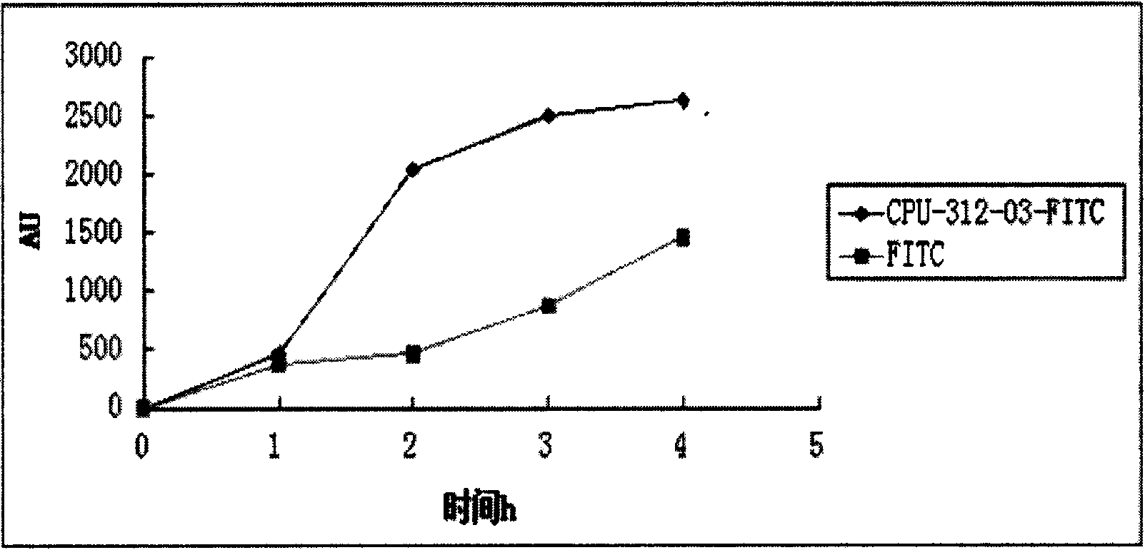
[0042] Take HepG2 cells (liver cancer cells) in the logarithmic growth phase, digest with an appropriate amount of 0.25% trypsin solution, and shake gently to make the digestive juice evenly act on the cells. RPMI-1640 medium with calf serum, blow gently with a pipette to form a single cell suspension; add 8×10 4 Put the cells in a 24-well plate, add 500 μl RPMI-1640 medium containing 10% serum, and incubate at 37°C with 5% CO 2 Cultivate in a constant temperature incubator for 24 hours to the state of monolayer cells; 1h, 2h, 3h, 4h before the test, add 500μl of RPMI-1640 medium containing 10% calf serum, 50um coupled with FITC-binding peptide, each time Spot 3 replicate wells and place at 37°C, 5% CO 2 Cultured in a constant temperature incubator; at the same time, FITC and LO2 cells (normal liver cells) were used as negative controls, and the fluorescence was de...
PUM
 Abstract
Description
Claims
Application Information
 - R&D
- Intellectual Property
- Life Sciences
- Materials
- Tech Scout
- Unparalleled Data Quality
- Higher Quality Content
- 60% Fewer Hallucinations
Browse by: Latest US Patents, China's latest patents, Technical Efficacy Thesaurus, Application Domain, Technology Topic, Popular Technical Reports.
© 2025 PatSnap. All rights reserved.Legal|Privacy policy|Modern Slavery Act Transparency Statement|Sitemap|About US| Contact US: help@patsnap.com


