A breast cancer circulating tumor cell detection system and kit
A tumor cell and detection system technology, applied in the field of breast cancer circulating tumor cell detection system and kits, can solve the problems of low specificity, false negative, wrong diagnosis, etc., and achieve the effect of high reliability of results and high separation purity
- Summary
- Abstract
- Description
- Claims
- Application Information
AI Technical Summary
Problems solved by technology
Method used
Image
Examples
Embodiment 1
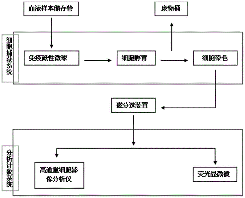
[0027] The circulating tumor cell detection instrument listed in this embodiment includes a cell capture system, an analysis and counting system, and a waste bin. The analysis and counting system includes a fluorescence microscope and a high-throughput cell image analyzer. The circulating tumor cell detection instrument is also equipped with Computer interface, through the computer to control the analysis and counting system.
[0028] Referring to Fig. 1, the workflow of the circulating tumor cell detection instrument is as follows: an optimized cell protection agent is placed in the blood sample storage tube, and the blood sample is collected and stored by using the blood sample storage tube; The antibody has immunomagnetic microbeads made of homologous secondary antibody magnetic beads, and the sample blood in the blood sample storage tube is placed in the functional enrichment box of the cell capture system, and the immunomagnetic microbeads and the target cells in the sampl...
Embodiment 2
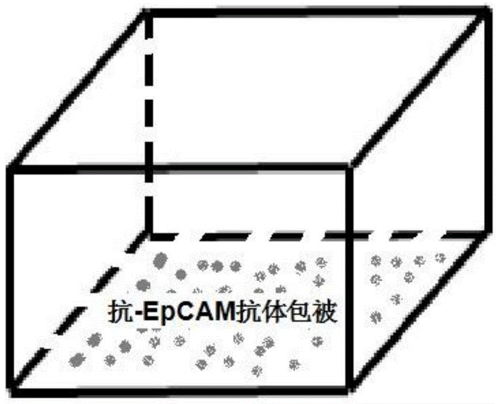
[0032] The use process of the functional enrichment box of the cell capture system
[0033] Open the functional enrichment package, the size is 10cm 3 , draw 7.5ml of peripheral blood sample with a syringe equipped with a sampling needle, anticoagulate with 3.8% sodium citrate 1 / 10 volume and centrifuge for 10min, 3000 rpm; draw the white blood cell layer in the middle, and put an equal volume of sterile double distilled water at room temperature Let stand for 5 minutes, centrifuge for 20 minutes; wash off the hemolyzed body in the supernatant, add an equal amount of normal saline, and centrifuge for 15 minutes; aspirate and discard the supernatant, add an equal amount of normal saline, centrifuge for 15 minutes, and save the obtained cells. Use a 1.0ml syringe to draw 0.5ml immunomagnetic microsphere sample, add it to a functional enrichment tube for incubation; after incubation for 30min, stain the cells, add phosphate buffer solution (pH=7.4, 0.1mol / L) to wash for 1-3 time...
Embodiment 3
[0035] Immunostaining analysis of tumor cells
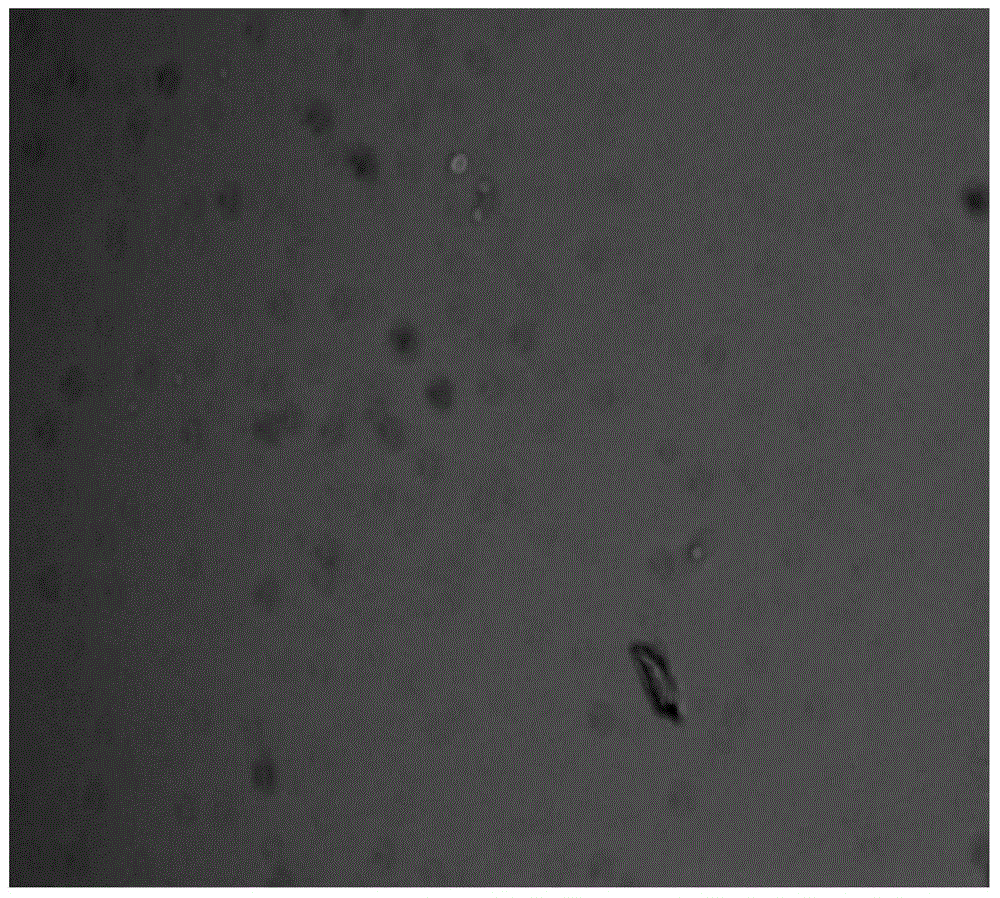
[0036] Blank magnetic balls and immunomagnetic balls loaded with Epcam antibody were used to "capture" breast cancer tumor cells for immunostaining, and observed under a fluorescent microscope, as shown in image 3 and Figure 4 Shown:
[0037] It can be seen from the figure that the ability of the immune magnetic ball loaded with Epcam antibody to capture breast cancer cells is significantly higher than that of the blank magnetic ball.
PUM
| Property | Measurement | Unit |
|---|---|---|
| particle diameter | aaaaa | aaaaa |
Abstract
Description
Claims
Application Information
 - R&D
- Intellectual Property
- Life Sciences
- Materials
- Tech Scout
- Unparalleled Data Quality
- Higher Quality Content
- 60% Fewer Hallucinations
Browse by: Latest US Patents, China's latest patents, Technical Efficacy Thesaurus, Application Domain, Technology Topic, Popular Technical Reports.
© 2025 PatSnap. All rights reserved.Legal|Privacy policy|Modern Slavery Act Transparency Statement|Sitemap|About US| Contact US: help@patsnap.com


