
PET and CT image lung tumor segmenting method based on graph cut

A CT image, lung tumor technology, applied in the field of biomedical image processing, can solve the problem of low accuracy and reliability

Inactive Publication Date: 2014-07-23
SUZHOU UNIV
View PDF2 Cites 19 Cited by
  • Summary
  • Abstract
  • Description
  • Claims
  • Application Information

AI Technical Summary

Problems solved by technology

These methods basically use a single modality to segment lung tumors, and the accuracy and reliability are not high.

Method used


Image

  • PET and CT image lung tumor segmenting method based on graph cut
  • PET and CT image lung tumor segmenting method based on graph cut
  • PET and CT image lung tumor segmenting method based on graph cut

Examples


Embodiment Construction

[0065] The present invention will be further described below in conjunction with the accompanying drawings.

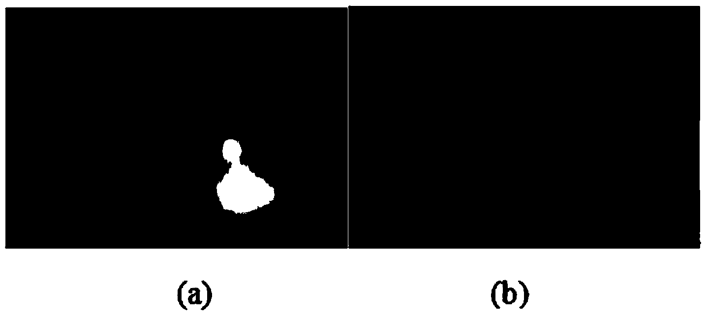
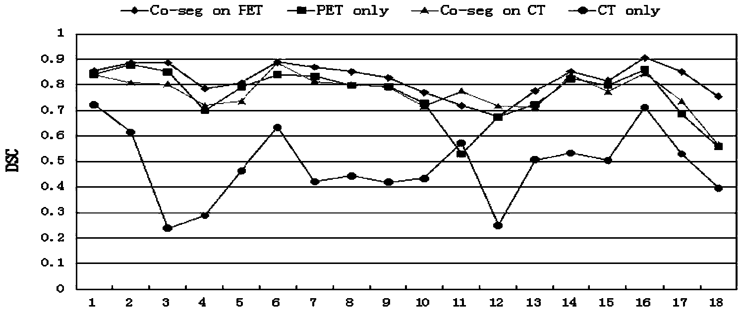
[0066] Such as figure 1 and figure 2 As shown, the lung tumor segmentation method of the present invention first collects PET and CT image data, and performs up-sampling on the PET image, and performs affine registration on the PET and CT images, so that the pixels on the PET and CT images are one by one Correspondence; calibrate the seed point of the tumor part and non-tumor part of the image; obtain the gold standard of the tumor with the help and supervision of clinical oncologists; extract the information of PET and CT, and use the algorithm of Graph cut to integrate analysis and extraction Based on the information on the PET and CT images, the lung tumor is segmented, tested, and the final detection result is obtained.

[0067] The method of the present invention is to obtain the data of patients with non-small cell lung cancer under the sponsorship of the Firs...


PUM


Abstract

The invention discloses a PET and CT image lung tumor segmenting method based on graph cut. The method includes the steps that first, PET image data acquisition and CT image data acquisition are conducted, a PET image is sampled, affine alignment is carried out on the PET image and a CT image, and accordingly pixels on the PET image and pixels on the CT image are made to be in one-to-one correspondence; seed point calibration is conducted on tumor locations and non-tumor locations of the images; a tumor golden standard of tumors is obtained with the help and supervision of clinical oncologists; through PET information extraction and CT information extraction, a lung tumor is segmented and tested by confluence analysis of the extracted information of the PET image and the CT image through a graph cut algorithm, and consequently a final testing result can be obtained.

Description

technical field [0001] The invention relates to a lung tumor segmentation method based on graph cut PET and CT images, which belongs to the field of biomedical image processing and uses a graph cut (graph cut) method to automatically optimize the segmentation method for lung tumors. Background technique [0002] At present, in today's deteriorating environment, lung tumors have become the main cause of harm to human health. The treatment of lung tumors requires accurate tumor location, size, and shape, so accurate lung tumor segmentation has become a hot research topic. As a quantitative molecular-structural imaging technique, PET-CT has been widely used in tumor analysis and formulation of tumor treatment plan. The optimal segmentation algorithm technology of Graph cut mainly combines the metabolic information of the human body provided by PET (positron emission tomography) images and the anatomical structure information provided by CT (computed tomography) to solve the se...

Claims


Application Information

Patent Timeline
IPC IPC(8): G06T7/00A61B6/03
Inventor 陈新建鞠薇章斌王振兴向德辉
Owner SUZHOU UNIV
Learn More