Kit for treating retinal disease

A technology for retinal diseases and kits, applied in the field of medical molecular biology, can solve problems such as retinal infection, hindering the dedifferentiation and proliferation of Müller cells, and the short-term effect of exogenous nutritional factors

Inactive Publication Date: 2014-09-03
THE FIRST AFFILIATED HOSPITAL OF THIRD MILITARY MEDICAL UNIVERSITY OF PLA
View PDF2 Cites 2 Cited by
  • Summary
  • Abstract
  • Description
  • Claims
  • Application Information

AI Technical Summary

Problems solved by technology

[0006] But, the shortcoming of this kind of method is: the effect of exogenous trophic factor is short-lived, if repeat subretinal cavity or vitreous cavity injection, easily cause intraocular infection, serious complications such as retinal infection, detachment, and Whether the rescued photoreceptors are still functional remains a question
Tip: The high expression of Let-7e and Let-7i may hinder the dedifferentiation and proliferation of Müller cells in the early stage of retinal degeneration

Method used


Image

  • Kit for treating retinal disease
  • Kit for treating retinal disease
  • Kit for treating retinal disease

Examples


Embodiment 1

[0056] Example 1, Preparation of recombinant adenovirus overexpressing RNA-binding protein Lin28B

[0057] Step (1) Preparation of Lin28B-encoding gene

[0058] Human Lin28B coding gene sequence: (GenBank: DQ127228.1)

[0059] Rat Lin28B coding gene sequence: (NCBI Reference Sequence: XM_001069344.2)

[0060] Step (2) Prepare the adenovirus shuttle vector pAV-MCMV-Lin28B-GFP-3FLAG

[0061] Include the following main steps:

[0062] The target gene was amplified by PCR using the chemically synthesized LIN28B gene as a template.

[0063] After digestion of the expression vector, the vector fragment was recovered by gel.

[0064] Transform E.coli competent cells after homologous recombination between the target gene and the vector fragment.

[0065] The transformants were identified by colony PCR, the positive clones were sent for sequencing, and the sequence verification was correct as the adenovirus shuttle vector pAV-MCMV-Lin28B-GFP-3FLAG. Clones were subjected to plasmi...

Embodiment 2

[0186] Embodiment 2, the formula of buffer solution and preparation method

[0187] The formula of the buffer solution is: 3% sucrose is dissolved in 1×PBS, that is, 3 g of sucrose is weighed and dissolved in 100 ml of 1×PBS, and sterilized by filtering through a 0.2 μm filter.

[0188] The preparation method of 1×PBS is as follows: 8g NaCl, 0.2g KCl, 1.44g Na2HPO4, 0.24g KH2PO4 are dissolved in 1 liter of distilled water, the pH value is 7.4, and autoclaved.

Embodiment 3

[0189] Embodiment 3, in vitro experiment:

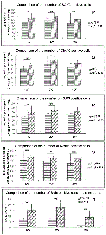
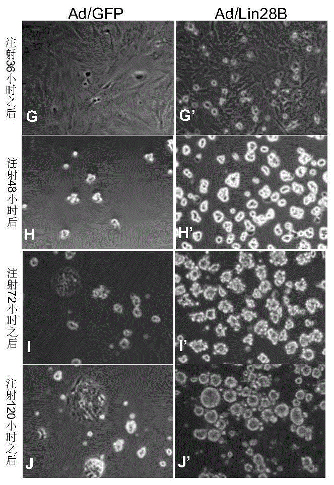
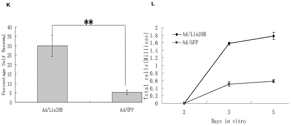
[0190] 1. In vitro observation of Ad / Lin28B on the self-renewal of Müller cells

[0191] 1) Prepare Müller cells infected with Ad / Lin28B.

[0192] Primary Müller Cell Culture Method

[0193] 1. Take a rat (LE mouse species, 7-10 days after birth, 8 days is the best) and place both eyes in Muller cell culture medium overnight (the time should not be too long, and should be controlled within 12 hours).

[0194] 2. Take out the overnight eyeball tissue and put it in the digestion solution (collagenase 0.5+trypsin 0.25) in a 37-degree incubator for 1 hour, and the muller medium stops the digestion.

[0195] 3. Peel off the retinal tissue on ice under a microscope, wash it once with medium at 4 degrees, and transfer it into an EP tube.

[0196] 4. Add muller cell culture medium into the EP tube and pipette not less than 100 times, gently and with few bubbles.

[0197] 5. After the pipetting is completed, balance it into the centrifuge...


PUM


Abstract

The invention discloses a kit for treating a retinal disease. The kit and a medicament for treating the retinal disease, which are disclosed by the invention, are characterized in that the effective component is adenovirus capable of carrying out overexpression on ribonucleic acid binding protein Lin28B. Injection for a plurality of times is not required when the retinal disease is treated by adopting the kit disclosed by the invention, retinal endogenous stem cells can be activated to the maximal extent by retina inferior vena injection, and the endogenous stem cells are promoted to be differentiated into retinal neurons.

Description

technical field [0001] The invention relates to the field of medical molecular biology, in particular to a kit for treating retinal diseases. Background technique [0002] Any eye disease that affects the normal function of the retina is called retinal disease, such as retinitis pigmentosa (RP), other inherited retinal degenerations, diabetic retinopathy (DR), retinal vessel occlusion, retinal detachment, retinal tear, age Related macular degeneration (AMD), etc. These diseases can affect the processing of visual information, and significantly affect the patient's visual function, severe cases can lead to blindness. Among them, wet and dry age-related macular degeneration is the leading cause of blindness in the elderly in developed countries, which is caused by the dysfunction of retinal pigment epithelium (RPE). In addition, the hereditary retinal degeneration represented by RP is mainly characterized by the destruction of photoreceptor cells and RPE, and the end result ...

Claims


Application Information

Patent Timeline
Patent Type & Authority Applications(China)
IPC IPC(8): C12N7/01C12N15/861A61K35/76A61K9/10A61P27/02A61P9/10
Inventor 李瑶琛阴正勤
Owner THE FIRST AFFILIATED HOSPITAL OF THIRD MILITARY MEDICAL UNIVERSITY OF PLA
Why Eureka
Try Eureka