Dual-mode imaging nano-micelle as well as preparation method and application thereof

A dual-mode imaging and nanomicelle technology, which is applied in the field of biomedical imaging, can solve the problems of complex preparation methods, high toxicity of bismuth sulfide, and unclear dual-mode imaging probe action duration and optimal imaging time, etc., to achieve high sensitivity , high spatial resolution, and reduced fluorescence quenching effects

Active Publication Date: 2014-10-29
THE NAT CENT FOR NANOSCI & TECH NCNST OF CHINA
View PDF2 Cites 7 Cited by
  • Summary
  • Abstract
  • Description
  • Claims
  • Application Information

AI Technical Summary

Problems solved by technology

However, the bismuth sulfide used in this patent is highly toxic, and the preparation method is complicated, and the duration of action of the dual-mode imaging probe and the optimal imaging time of the two modes are not clear

Method used


Image

  • Dual-mode imaging nano-micelle as well as preparation method and application thereof
  • Dual-mode imaging nano-micelle as well as preparation method and application thereof
  • Dual-mode imaging nano-micelle as well as preparation method and application thereof

Examples


Embodiment 1

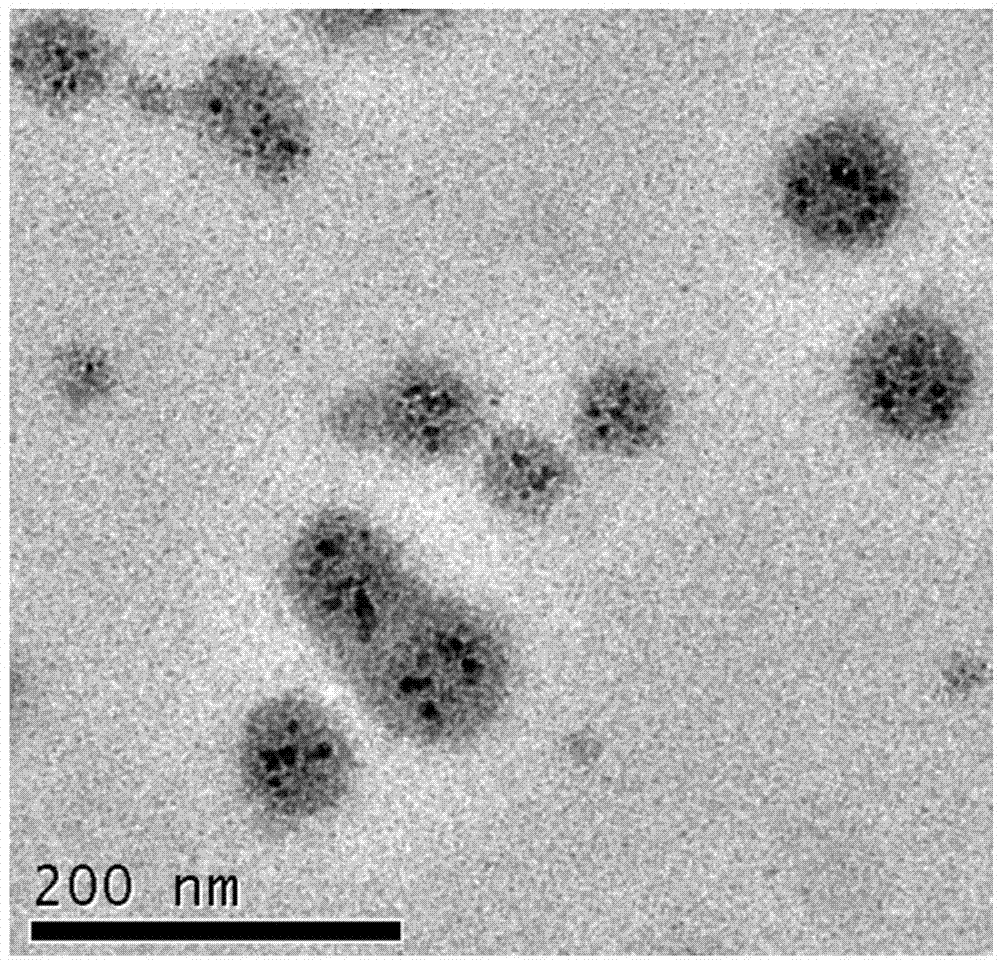
[0039] Microscopic morphological observation of the dual-mode imaging nanomicelles of the present invention:

[0040]Electron microscope scanning is performed on the dual-mode imaging nanomicelles prepared by the present invention, see figure 1 , it can be seen that the dual-mode imaging nanomicelles are spherical particles with a diameter of about 80 nm.

Embodiment 2

[0042] The dual-mode imaging nanomicelle of the present invention is prepared by the following method:

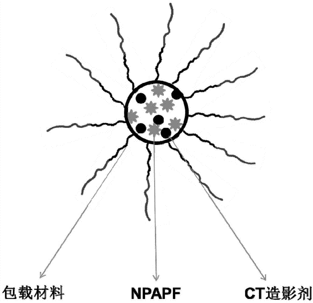
[0043] Dissolve CT imaging agent, NPAPF and PLGA-PEG in chloroform in a mass ratio of 1:1:8;

[0044] The obtained chloroform solution was mixed with 10 times the volume of deionized water, and the organic solvent chloroform was removed to obtain the dual-mode imaging nanomicelles.

Embodiment 3

[0046] The dual-mode imaging nanomicelle of the present invention is prepared by the following method:

[0047] Dissolve CT imaging agent: NPAPF: DSPE-PEG in chloroform in a mass ratio of 1:1:8;

[0048] The obtained chloroform solution was mixed with 10 times the volume of deionized water, and the organic solvent chloroform was removed to obtain the dual-mode imaging nanomicelles.


PUM


Abstract

The invention discloses dual-mode imaging nano-micelle which comprises a CT (Computed tomography) contrast medium, red fluorescent molecules NPAPF (N-(2-naphthyl)phenylamino)phenyl)-fumaronitrile) and a carrying material for carrying the CT contrast medium and the NPAPF into the dual-mode imaging nano-micelle. The invention further discloses a preparation method and an application of the dual-mode imaging nano-micelle. The dual-mode imaging nano-micelle combines fluorescent imaging and CT imaging, achieves targeted dual-mode imaging of living tumor and organs and meanwhile is long in action time, intensive in detection signal and very small in toxicity.

Description

technical field [0001] The invention belongs to the field of biomedical imaging, relates to a nanomicelle, a preparation method and application thereof, and more particularly relates to a dual-mode imaging nanomicelle, a preparation method and application thereof, which combine fluorescence imaging and CT imaging , enabling in vivo targeting and imaging of animals. Background technique [0002] Malignant tumor is one of the most important diseases endangering human health. According to the data released by the International Union Against Cancer, in 2008, 12.7 million people worldwide had cancer and 7.6 million died. Without effective measures, it is estimated that by 2030, there will be 26 million new cancer cases each year. Cancer has become the number one cause of death for human beings both in the world and in China. Therefore, the development of effective cancer imaging diagnostic agents is of great significance to promote the health of Chinese people. [0003] Aggre...

Claims


Application Information

Patent Timeline
Patent Type & Authority Applications(China)
IPC IPC(8): A61K49/00A61K49/04
Inventor 梁兴杰张吉梅安菲菲李婵李鸿基王东亮张旭霍帅东金叔宾姜永刚
Owner THE NAT CENT FOR NANOSCI & TECH NCNST OF CHINA
Why Eureka
Try Eureka