Tissue microstructure detection method based on empirical mode decomposition in quantitative ultrasound system

An empirical mode decomposition and detection method technology, which is applied in the fields of ultrasonic/sonic/infrasonic Permian technology, organ movement/change detection, ultrasonic/sonic/infrasonic image/data processing, etc. Scattering signal noise and diffuse scattering signal interference, etc., to eliminate signal and noise interference, suppress interference peaks, and improve estimation accuracy

Active Publication Date: 2014-11-12
哈尔滨工业大学人工智能研究院有限公司
View PDF6 Cites 9 Cited by
  • Summary
  • Abstract
  • Description
  • Claims
  • Application Information

AI Technical Summary

Problems solved by technology

[0005] In order to solve the problem that the noise in the ultrasonic backscattering signal and the interference of the diffuse scattering signal cannot be effectively eliminated by using the prior art methods in the quantitative ultrasound system, the present invention provides a tissue microscopic method based on Empirical Mode Decomposition (EMD) in the quantitative ultrasound system. Structural detection method

Method used


Image

  • Tissue microstructure detection method based on empirical mode decomposition in quantitative ultrasound system
  • Tissue microstructure detection method based on empirical mode decomposition in quantitative ultrasound system
  • Tissue microstructure detection method based on empirical mode decomposition in quantitative ultrasound system

Examples


Embodiment Construction

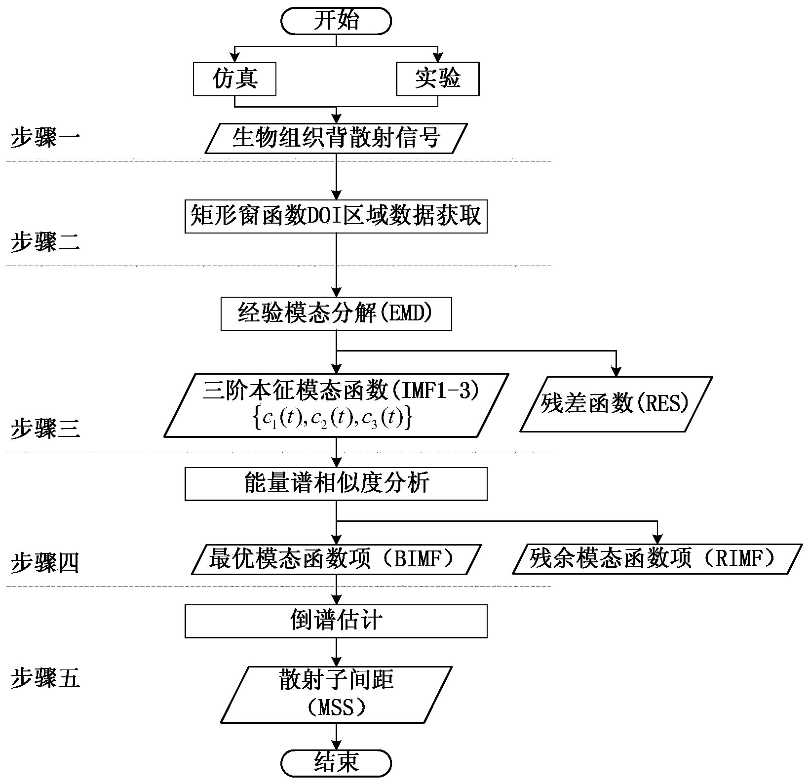
[0042] The technical solution of the present invention will be further described below in conjunction with the accompanying drawings, but it is not limited thereto. Any modification or equivalent replacement of the technical solution of the present invention without departing from the spirit and scope of the technical solution of the present invention should be covered by the present invention. within the scope of protection.

[0043] The present invention provides a numerical simulation method for internal stress distribution of bone tissue based on finite element ultrasonic physiotherapy, and its realization process is as follows: figure 1 As shown, it specifically includes the following steps:

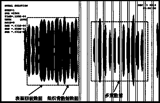
[0044] (1) Obtain ultrasonic backscattering signals of biological tissues.

[0045] There are two methods of obtaining ultrasonic backscattering signals of biological tissues including simulation and experiment.

[0046] The simulation data acquisition method adopts the finite ele...


PUM


Abstract

The invention discloses a tissue microstructure detection method based on empirical mode decomposition in a quantitative ultrasound system. The detection method comprises the steps that firstly, biological tissue ultrasound back-scattered signals are obtained; secondly, the back-scattered signals in a DOI are intercepted to serve as input data; thirdly, EMD is conducted on the input data so as to obtain a three-order intrinsic mode function and a residual term; fourthly, the energy spectrum of the three-order intrinsic mode function is extracted, similarity analysis is conducted on the energy spectrum of the three-order intrinsic mode function and the energy spectrum of the original signals, and a BIMF containing tissue microstructure information is determined; fifthly, the determined BIMF is estimated, and tissue scatterer spacing information is worked out. The method has the advantages that the BIMF is extracted, and useless information in non-coherent mode function items is removed; signals and noise interference of dispersion scatterers are eliminated, an interference peak value in a cepstrum is restrained, and the estimation accuracy of MSS is improved and can reach 90%-95%.

Description

technical field [0001] The invention relates to a method for detecting the average scattering sub-distance characterizing tissue microstructure by using ultrasonic backscattering technology. Background technique [0002] Medical ultrasound is widely used in human tissue and organ imaging. Although this technology has a fairly mature application basis in clinical applications and can provide real-time high-precision human organ images, it cannot provide microscopic images caused by tissue calcification and variation. Structural changes. [0003] At present, some equivalent scattering sub-models such as binary mixture model, Faran cylinder model and weak scattering sub-model are commonly used to characterize tissue microstructure changes. In these models, the detection of tissue is transformed into the detection of ultrasonic attenuation, backscatter coefficient, scatterer density, scatterer diameter and mean scatterer spacing (MSS), etc. Among them, MSS is the most commonly...

Claims


Application Information

Patent Timeline
Patent Type & Authority Applications(China)
IPC IPC(8): A61B8/08
Inventor 沈毅潘文磊金晶王艳章欣
Owner 哈尔滨工业大学人工智能研究院有限公司
Why Eureka
Try Eureka