Passage method and application of human induced pluripotent stem cells
A pluripotent stem cell and cell technology, applied in the field of single cell passage and culture of human induced pluripotent stem cells, can solve the problems of difficulty in large-scale expansion, easy differentiation of hiPSCs, low efficiency of cryopreservation and recovery, and achieve the effect of improving efficiency.
- Summary
- Abstract
- Description
- Claims
- Application Information
AI Technical Summary
Problems solved by technology
Method used
Image
Examples
Embodiment 1
[0040] Example 1 hiPSCs single cell passage
[0041] 1) Spread ECM on a 6-well plate 1-2 hours before cell subculture.
[0042] 2) Take out the cells with a confluence rate of about 75%-85% from the incubator, wash once with DMEM / F12 medium, add Accutase digestive enzyme and place in 37°C with 12% volume fraction of CO 2 Digest in the incubator, when the cells are basically digested, add mTeSR1 to stop the digestion.
[0043] 3) Centrifuge at 200g for 3 minutes, discard the supernatant, resuspend the cells in mTeSR1 containing Y27632, and gently pipette evenly.
[0044] 4) Count the cells with a hemocytometer, inoculate them on a 6-well plate pre-coated with ECM at a density of 20,000-30,000 / cm2, and place them in CO at 37°C and a volume fraction of 12%. 2 Incubator cultivation.
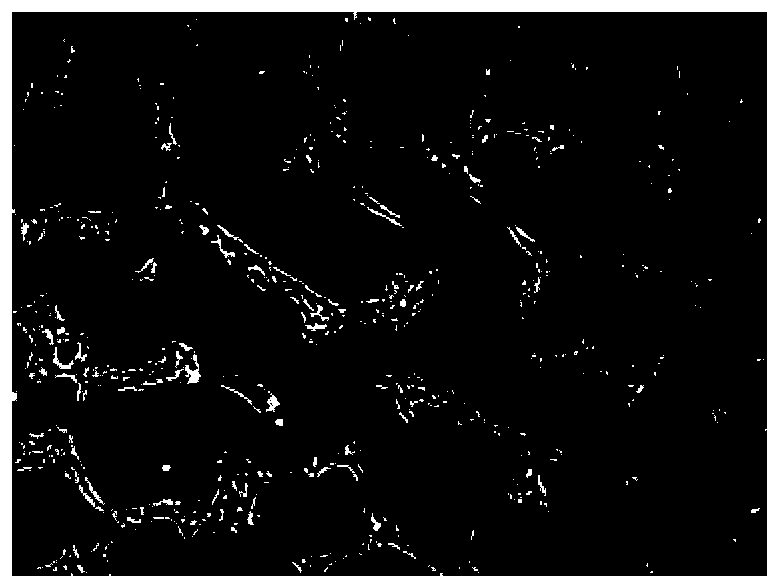
[0045] 5) The next day, observe the cell density and adhesion under the microscope ( Figure 1A ). If there is contact between the cells and the cell density is appropriate, remove Y27632 and repla...
Embodiment 2
[0048] Example 2 hiPSCs single cell clone
[0049] 1) Take out cells with a confluence rate of about 75%-85% from the incubator, add Accutase digestive enzyme and place in 37°C, 8-15% volume fraction of CO 2 Digest in the incubator, when the cells are basically digested, add mTeSR1 to stop the digestion.
[0050] 2) Trypan blue staining, use a hemocytometer to count the number of viable cells, use mTeSR1+Y27632 inhibitor to dilute the cell suspension to 5-15 cells / ml, add 100ul of the cell suspension to the ECM-coated 96-well plate / Well, put in 8-15% CO 2 cultured in an incubator.
[0051] 3) After 10 days of microscopic examination, the cloned hiPSCs were passaged and expanded to obtain single-cell-derived hiPSCs.
Embodiment 3-h
[0052] Example 3- Identification of pluripotency after single-cell passage of hiPSCs
[0053] After hiPSCs were single-cell passaged for 20 passages using the method in Example 1, some cells were taken for EB formation experiments and Teratomas formation experiments.
[0054] 1) Embryooid body formation
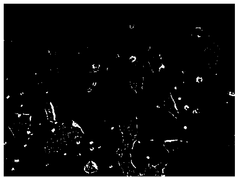
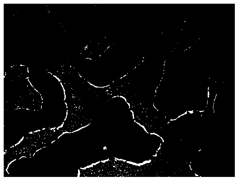
[0055] Resuscitate hiPSCs on the ECM, and transfer the cells to the feeder for culture when the cells grow to 75%. After the clonal group grows on the feeder for 5-6 days, transfer the clonal group to suspension culture ( Figure 2A ). After 8 days of suspension culture, EB balls were transferred to gelatin-coated 6-well plates for adherent growth. After 8 days of EB ball adherent growth, the RNA of adherent cells was collected, and the expression of three germ layer markers was detected by qPCR ( Figure 2B ). The results showed that compared with hiPSCs, the cells differentiated by EBs increased in the three germ layer cell markers, and compared with hiPSCs, there was ...
PUM
 Abstract
Description
Claims
Application Information
 - R&D
- Intellectual Property
- Life Sciences
- Materials
- Tech Scout
- Unparalleled Data Quality
- Higher Quality Content
- 60% Fewer Hallucinations
Browse by: Latest US Patents, China's latest patents, Technical Efficacy Thesaurus, Application Domain, Technology Topic, Popular Technical Reports.
© 2025 PatSnap. All rights reserved.Legal|Privacy policy|Modern Slavery Act Transparency Statement|Sitemap|About US| Contact US: help@patsnap.com


