Lung respiration monitoring system based on magnetic detection electrical impedance imaging

A respiratory monitoring system and electrical impedance imaging technology, applied in the field of biomedical imaging, can solve the problems of low image resolution and less measurement information, and achieve the effect of improving reconstruction quality, improving morbidity, and improving the limitation of measurement information

Inactive Publication Date: 2015-07-22
TIANJIN POLYTECHNIC UNIV
View PDF5 Cites 12 Cited by
  • Summary
  • Abstract
  • Description
  • Claims
  • Application Information

AI Technical Summary

Problems solved by technology

[0010] The purpose of the present invention is to provide a lung respiration monitoring system based on magnetic detection electrical impedance imaging, which solves the problems of less measurement information and low image resolution in the background technology. See the following description for details:

Method used


Image

  • Lung respiration monitoring system based on magnetic detection electrical impedance imaging
  • Lung respiration monitoring system based on magnetic detection electrical impedance imaging
  • Lung respiration monitoring system based on magnetic detection electrical impedance imaging

Examples


Embodiment 1

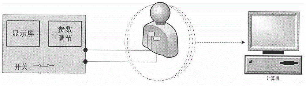
[0040] see image 3 , the excitation source in the respiratory monitoring system is a current source with a certain frequency and intensity. The current excitation signal is stimulated to the measured object through the electrode array pasted around the chest of the human body, and a magnetic field will be generated around the chest; the magnetic field information at different positions can be obtained through the coils placed around the measured object; finally, the image reconstruction algorithm is used in the computer The distribution images of the electrical impedance-sensitive areas inside the thorax during respiration are obtained on the above image. When performing respiratory monitoring, the subject sits on a chair, places the excitation electrodes on the imaging body, connects the measuring coil to the monitoring chair through a bracket of a certain shape, and fixes it around the chest cavity of the imaging body The excitation and measurement process is controlled by...

Embodiment 2

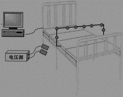
[0042] see Figure 4 , the excitation source in the respiratory monitoring system is a voltage source with a certain frequency and strength. The voltage excitation signal is stimulated to the measured object through the electrode array pasted around the chest of the human body, and a magnetic field will be generated around the chest; the magnetic field information at different positions can be obtained through the coils placed around the measured object; finally, the image reconstruction algorithm is used in the computer The distribution images of the electrical impedance-sensitive areas inside the thorax during respiration are obtained on the above image. When performing respiratory monitoring, the subject lies on his back on the detection bed, the excitation electrodes are placed on the subject, the measuring coil is connected to the monitoring bed through a bracket of a certain shape, and fixed around the chest of the subject, controlled by the control module The process o...


PUM


Abstract

The invention discloses a lung respiration monitoring system based on magnetic detection electrical impedance imaging. By means of an excitation source module, excitation signals are introduced to the chest cavity of an imaging body through a plurality of pairs of excitation electrodes bonded to the measured imaging body; a measurement module is composed of a series of measurement coils and used for measuring the magnetic induction intensity of an induced magnetic field caused by exciting current around the chest cavity; a control module is used for controlling the signal excitation and magnetic field measurement process during the whole respiration monitoring; an image reconstructing module obtains the image change of electrical inductance distribution of the chest cavity in the respiration process according to an electrical impedance image reconstruction algorithm on the basis of magnetic field distribution data. According to the system, the measurement coils of any number can be placed in the space around the chest cavity of the imaging body, limitation of a traditional electrical impedance imaging system on measurement information is changed, the system has the advantages of being high in measurement precision, high in speed and reliability and free of injuries to a human body, more importantly, information amount is increased by means of a magnetic field measurement mode, and therefore the electrical impedance reconstructing ill conditioning is changed, the resolution ratio of electrical impedance (or impedance spectrum) images is increased, and the image reconstruction quality is improved.

Description

technical field [0001] The invention relates to the field of biomedical imaging, in particular to a lung respiration monitoring system based on magnetic detection electrical impedance imaging. Background technique [0002] Electrical impedance tomography is a non-invasive functional imaging method that can be used for real-time monitoring of the respiratory process. patent [1] The three-dimensional electrical impedance imaging system built with the help of the highly integrated hardware and software tool suite of the PXI bus has the advantages of fast speed, no damage to the human body, and continuous real-time bedside monitoring. In addition, an electrical impedance imaging system for respiratory monitoring designed by Hebei University of Technology [2] , using direct digital synthesizer to build multi-frequency digital excitation signal source, using DSP as the core device of system control and processing, and perfecting each functional module of the system. EIT_TJU sys...

Claims


Application Information

Patent Timeline
Patent Type & Authority Applications(China)
IPC IPC(8): A61B5/08A61B5/053
CPCA61B5/0809A61B5/0536A61B5/6891A61B5/7221
Inventor 陈瑞娟尹剑利王金海王慧泉
Owner TIANJIN POLYTECHNIC UNIV
Why Eureka
Try Eureka