FCN (Fluorescent carbon nano-particle) based visual spherical nucleic acid as well as preparation method and application thereof

A carbon nanoparticle and nucleic acid technology, applied in the visualization of spherical nucleic acid and its preparation, based on fluorescent carbon nanoparticles in the field of visualization of spherical nucleic acid and its preparation, can solve the problems of limited application range of FCNs, and achieve easy scale and promotion, high efficiency Delivery to, low cost effect

Active Publication Date: 2015-07-29
THE NAT CENT FOR NANOSCI & TECH NCNST OF CHINA
View PDF4 Cites 0 Cited by
  • Summary
  • Abstract
  • Description
  • Claims
  • Application Information

AI Technical Summary

Problems solved by technology

However, the application range of FCNs is relatively limited at present, and how to

Method used


Image

  • FCN (Fluorescent carbon nano-particle) based visual spherical nucleic acid as well as preparation method and application thereof
  • FCN (Fluorescent carbon nano-particle) based visual spherical nucleic acid as well as preparation method and application thereof
  • FCN (Fluorescent carbon nano-particle) based visual spherical nucleic acid as well as preparation method and application thereof

Examples


Embodiment 1

[0046] Embodiment 1: Preparation and separation and purification of fluorescent carbon nanoparticles (FCNs)

[0047] (1) Preparation of fluorescent carbon nanoparticles (FCNs):

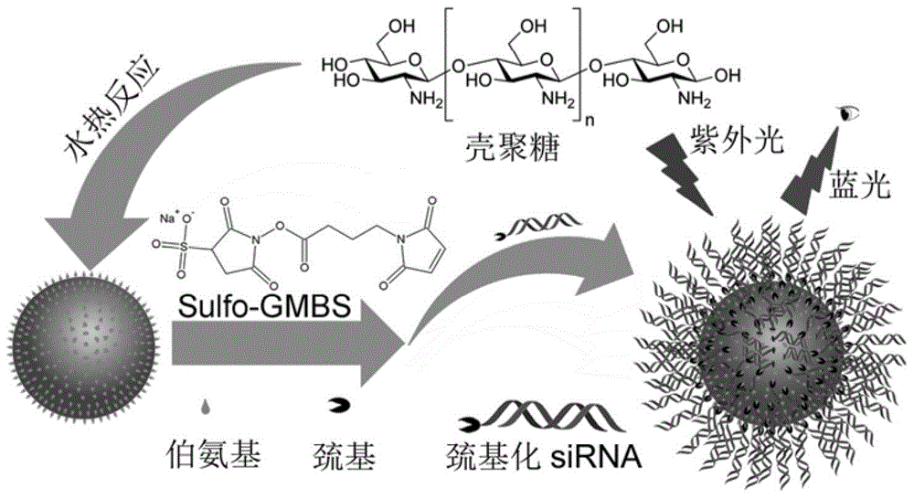
[0048] Using low-molecular-weight chitosan as a raw material, carbon nanoparticles with good fluorescence characteristics were obtained by hydrothermal synthesis. The preparation method was: weigh 0.35 g of low-molecular-weight chitosan and add it to 70 mL of deionized water while fully stirring. Then place it in an airtight container (autoclave) for 12 hours of reaction at a temperature of 180°C.

[0049] (2) Separation and purification of fluorescent carbon nanoparticles (FCNs):

[0050] The resulting product solution was centrifuged in a high-speed desktop refrigerated centrifuge (14000 rpm) for 10 min, and then treated with a dialysis bag (molecular weight cut-off 1000 Da) to remove small molecular substances, and the dialysis time was 2 days.

Embodiment 2

[0051] Embodiment 2: Characterization of properties of fluorescent carbon nanoparticles (FCNs)

[0052] (1) Fourier infrared spectroscopy of fluorescent carbon nanoparticles

[0053] figure 2 A is the infrared spectrogram of fluorescent carbon nanoparticles, composed of figure 2 A It can be seen that the fluorescent carbon nanoparticles prepared in Example 1 have an N-H stretching vibration absorption peak at around 1660-1580.

[0054] (2) High-definition transmission electron microscope characterization of fluorescent carbon nanoparticles

[0055] figure 2 B is the high-definition transmission electron microscope characterization diagram of fluorescent carbon nanoparticles, by figure 2 B, it can be seen that the particle size of fluorescent carbon nanoparticles is 11.83±2.22nm, and the particle size distribution is relatively uniform, and the monodispersity is good.

[0056] (3) Imaging of fluorescent carbon nanoparticles

[0057] figure 2 C is the picture of fluo...

Embodiment 3

[0058] Example 3: Cytotoxicity Test of Fluorescent Carbon Nanoparticles (FCNs)

[0059] Put MDA-MB-231 and A375 cells into a 96-well plate at a density of 5000 / well in 5% CO 2 , and incubated in a 37°C incubator for 24h. Fluorescent carbon nanoparticles (FCNs) were added at concentrations of 0, 10, 20, 50, 80, 100, 150, 200, 250, 300 and 350 μg / mL, respectively. 5 replicate wells for each concentration. After 48 hours, suck out the culture medium, add and replace with 200 μL freshly prepared 3-(4,5-dimethylthiazole-2)-2,5-diphenyltetrazolium bromide at a concentration of 5 mg / ml in each well (MTT) solution, continue culturing for 4 hours, suck out the culture medium in the wells, add 200 μL of dimethyl sulfoxide to each well, shake on a shaker at low speed for 10 minutes, and fully dissolve the crystals. The absorbance of each well was measured at OD490nm in an enzyme-linked immunosorbent assay instrument.

[0060] Depend on image 3 It can be seen that the toxicity of th...


PUM


Abstract

The invention relates to FCN (Fluorescent carbon nano-particle) based visual spherical nucleic acid as well as a preparation method and an application thereof. The visual spherical nucleic acid comprises FCNs and oligonucleotide combined on the surfaces of the FCNs. The FCNs with good fluorescence characteristics are obtained with a one-step reaction method and then are combined with oligonucleotide to prepare a visual spherical nucleic acid system; according to the method, a preparation process is simplified, nucleic acid can be effectively delivered into a cell by the visual spherical nucleic acid under the condition of absence of any other transfection reagents, the intake rate and the transfection efficiency of the cell are increased, and important application value is achieved.

Description

technical field [0001] The invention relates to the technical field of biopharmaceuticals, in particular to a visualized spherical nucleic acid and its preparation method and application, in particular to a fluorescent carbon nanoparticle-based visualized spherical nucleic acid and its preparation method and application. Background technique [0002] Gene therapy has emerged as a novel and powerful approach to treating challenging diseases such as cancer. Effective gene therapy relies heavily on safe and efficient transfection reagents. However, delivery of therapeutic nucleic acids, including siRNA and antisense DNA, remains a major difficulty in gene regulation and therapy. Nucleic acid delivery vehicles, such as cationic polymers, liposomes and artificially engineered viruses, are used for the delivery of nucleic acids to achieve the purpose of penetrating cell membranes and protecting them from nuclease degradation. There are great deficiencies in these carriers, such ...

Claims


Application Information

Patent Timeline
IPC IPC(8): A61K48/00A61K31/7088A61K49/00C12Q1/68C12N15/11A61P35/00
Inventor 蒋兴宇张灵敏郑文富靳钰
Owner THE NAT CENT FOR NANOSCI & TECH NCNST OF CHINA
Why Eureka
Try Eureka