Choroidal vessel extraction method based on sd‑oct retinal images

A retinal and choroidal technology, applied in the field of target area extraction, can solve problems such as high complexity, difficult implementation, and given choroidal vascular area, so as to achieve the effect of improving efficiency

Inactive Publication Date: 2018-01-19
THE FIRST AFFILIATED HOSPITAL OF MEDICAL COLLEGE OF XIAN JIAOTONG UNIV
View PDF6 Cites 0 Cited by
  • Summary
  • Abstract
  • Description
  • Claims
  • Application Information

AI Technical Summary

Problems solved by technology

Because the choroidal vessels are not obvious on the SD-OCT image, it is difficult for the traditional target extraction method to effectively identify the choroidal vessel area. At present, there is only one document in the world that introduces a choroidal vessel extraction based on SD-OCT images. Method: The multi-scale Hessian matrix analysis method was used to obtain the structure tensor of each small cube of the choroidal layer, and then the choroidal vessels were extracted by the eigenvalues ​​of the structure tensor
This method is difficult to implement and has high complexity
[0003] Therefore, the practicability of the existing choroidal blood vessel extraction method is not high, and it is difficult to meet the needs of clinical ophthalmic disease diagnosis

Method used


Image

  • Choroidal vessel extraction method based on sd‑oct retinal images
  • Choroidal vessel extraction method based on sd‑oct retinal images
  • Choroidal vessel extraction method based on sd‑oct retinal images

Examples


Embodiment Construction

[0028] The present invention will be described in detail below in conjunction with the accompanying drawings and embodiments.

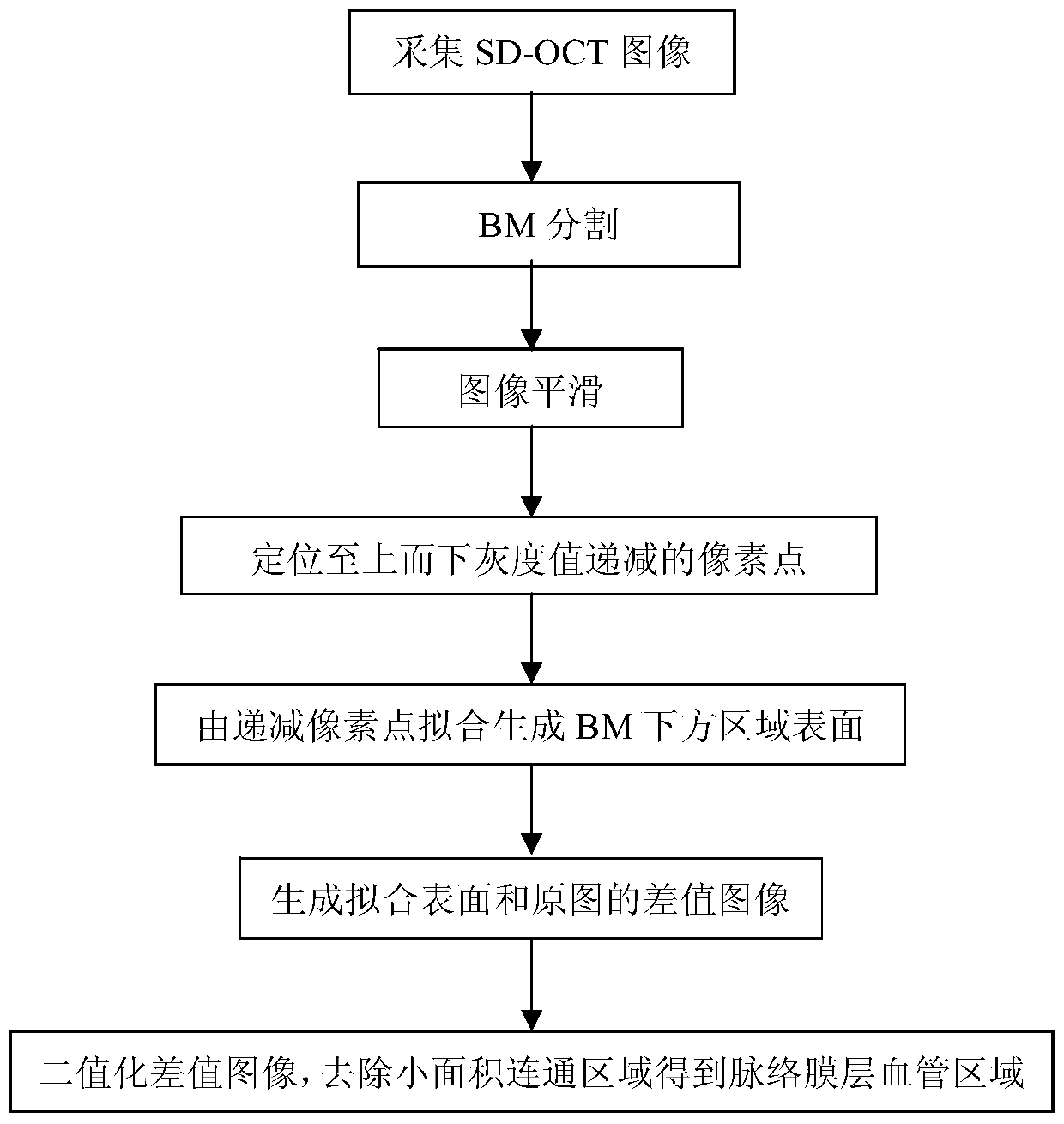
[0029] combine figure 1 The choroidal vessel extraction method based on the SD-OCT retinal image of the present invention uses the SD-OCT retinal image as an input, and uses image processing means to extract the choroidal vessel region, comprising the following steps:

[0030] Step 1, collecting SD-OCT retinal images, using existing OCT imaging equipment to collect retinal images;

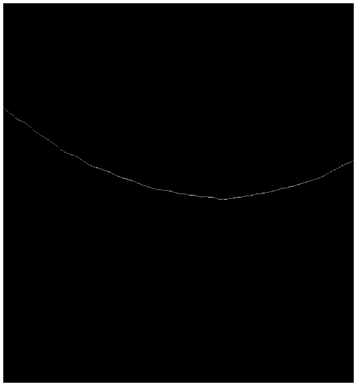
[0031] Step 2. Manually segment the BM boundary; figure 2 Given a frame of SD-OCT retinal image ( figure 1 , the size of the three-dimensional SD-OCT retinal image collected by OCT imaging equipment is 1024×512×128, corresponding to the area of ​​interest in the retinal area of ​​2mm×6mm×6mm), the white line in the figure is the boundary of BM (Bruch’s membrane) , which is the upper border of the choroid, and below the BM are the choroid and sclera regions;

[0032] S...


PUM


Abstract

The invention discloses a choroid membrane blood vessel extraction method based on an SD-OCT retina image, and belongs to the technical field of image processing. The method comprises the following steps: firstly performing Bruch's membrane partitioning on the input SD-OCT image, then carrying out smoothing by adopting bilateral filter, finding pixel points of which the grey scale values are monotonically decreased from top to bottom in each column in a regional image below the smoothed BM, fitting the monotonically decreased pixel points by cubic interpolation values based on a triangle to obtain a binary difference image which is positioned between a fitting surface of the region below the BM region and an original image, and finally removing a small-area communicating region in a binary result to obtain a blood vessel region of a choroid membrane layer. An experiment result shows that large blood vessels of the choroid membrane layer can be better extracted, and the choroid membrane blood vessel extraction method has great significance for facilitating the subsequent retinal disease analysis and improving the working efficiency of a doctor.

Description

technical field [0001] The invention relates to a method for extracting a target area, in particular to a method for extracting choroidal blood vessels based on a frequency-domain optical coherence tomography (SD-OCT) retinal image. Background technique [0002] SD-OCT retinal image is a frequency-domain optical coherence tomography image, which can effectively present changes in the grayscale and structure of retinal tissue layers. Clinical experiments have shown that SD-OCT images can also be used to image choroidal blood vessels. The choroid layer is composed of a large number of blood vessels, which provide nutrition for the retinal layer and is closely related to retinal diseases. Because the choroidal vessels are not obvious on the SD-OCT image, it is difficult for the traditional target extraction method to effectively identify the choroidal vessel area. At present, there is only one document in the world that introduces a choroidal vessel extraction based on SD-OCT i...

Claims


Application Information

Patent Timeline
Patent Type & Authority Patents(China)
IPC IPC(8): G06T7/10
CPCG06T7/12G06T2207/10101G06T2207/30041G06T2207/30101
Inventor 权芳吴昌睿刘利锋李白芽秦莉陈蕴光
Owner THE FIRST AFFILIATED HOSPITAL OF MEDICAL COLLEGE OF XIAN JIAOTONG UNIV
Why Eureka
Try Eureka