Application of p53 gene in reversing the malignant characteristics of oral mucosal malignant melanoma cells and its in vitro evaluation method

A malignant melanoma, p53 gene technology, applied in the field of cell and molecular biology, can solve the problems of no gene therapy method, imperfect characterization method, immature technology, etc.

Inactive Publication Date: 2018-06-19
李龙江
View PDF0 Cites 0 Cited by
  • Summary
  • Abstract
  • Description
  • Claims
  • Application Information

AI Technical Summary

Problems solved by technology

However, as an emerging cancer treatment method, gene therapy is not yet mature, and its inhibitory mechanism for different types of cancer is not clear, and the treatment strategies for different types of cancer are also different.
Especially for oral mucosal melanoma, there is no relatively mature gene therapy method at present, and the current characterization method for the reversal of the malignant characteristics of oral mucosal melanoma cells is not perfect

Method used


Image

  • Application of p53 gene in reversing the malignant characteristics of oral mucosal malignant melanoma cells and its in vitro evaluation method
  • Application of p53 gene in reversing the malignant characteristics of oral mucosal malignant melanoma cells and its in vitro evaluation method
  • Application of p53 gene in reversing the malignant characteristics of oral mucosal malignant melanoma cells and its in vitro evaluation method

Examples


Embodiment 1

[0047] Example 1 Passage and culture of human malignant melanoma cell line (A-375)

[0048] Take frozen A375 cells for recovery, add RPMI1640 (containing 10% calf serum) cell culture medium for culture, observe the cells, and passage when the culture bottle is 80% full.

[0049] figure 1 Photos of normal growth of A-375 cells entering the logarithmic growth phase under 100X and 200X microscopes. It can be seen from the figure that the cells grow well.

Embodiment 2

[0050] Example 2 Determination of the transfection efficiency of rAd-GFP to A-375

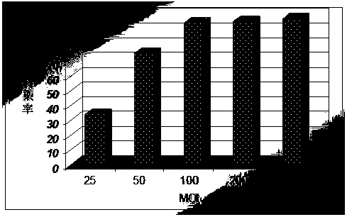
[0051] Take the A-375 cells recovered in Example 1, and use 1×10 5 The density of each well was inoculated in a 6-well culture plate for 24 hours after the cells adhered to the wall, the supernatant was sucked off, and the recombinant adenovirus (rAd-GFP) virus solution with the green fluorescent protein gene was used at MOI=0, 25, 50, 100, 200, 500 infected cells. After 48 hours, under a fluorescence microscope, randomly select three 200-field views in each well to count the number of GFP-positive cells showing green fluorescence, and at the same time count the total number of cells in the field of view under visible light, and calculate the transfection rate according to the formula (transfection rate r = number of GFP positive cells / total number of cells in the field of view).

[0052] The result is as figure 2 and image 3 As shown, 10-12 hours after transfection with Ad-GFP, A-375 cel...

Embodiment 3

[0053] Example 3 MTT method to detect the growth inhibition rate of A-375 cells under different rAd-p53 transfection intensities

[0054] Take the A-375 cells recovered in Example 1, and use 5×10 4 Inoculate each well in a 96-well plate, routinely culture for 24 hours after the cells adhere to the wall, absorb the culture medium, add recombinant human p53 adenovirus (rAd-p53) injection according to MOI=0, 25, 50, 100, 200, 500, Set rAd-p53 group and blank control group (only add complete culture medium), and set up three parallel wells for each concentration in each group.

[0055] MTT staining method was used to detect the growth inhibition rate of cells, and the optical density (OD) values ​​at 24, 48, 72, 96, and 120 hours were measured with a porous high-efficiency analyzer at a wavelength of 570 nm, and the cell proliferation inhibition was calculated. Rate. Proliferation inhibition rate=(OD value of control group-OD value of treatment group) / OD value of control group×1...


PUM


Abstract

The invention discloses the application of the p53 gene to reversing of mouth mucosa malignant melanoma cell malignant characteristics, and an in vitro evaluation method for reversing of the mouth mucosa malignant melanoma cell malignant characteristics, and belongs to the technical field of cellular and molecular biology. According to the invention, a gene therapy method is applied to the reversing of mouth mucosa malignant melanoma cell malignant characteristics and the in vitro evaluation method for reversing of the mouth mucosa malignant melanoma cell malignant characteristics; by introducing the exogenous wild-type p53 gene into a malignant melanoma-related cell strain, using various mechanisms, and depending on synthetic action of various signal transduction pathways, cell growth is inhibited, meanwhile, the cell cycle distribution is changed, cells are induced for apoptosis, and the malignant characteristics of cell transfer and invasion can be reversed. The evaluation method is simple and reliable, is scientific and rational, is capable of comprehensively detecting the reversing condition of the mouth mucosa malignant melanoma cell malignant characteristics, and can be used not only for studying of oral mucosa malignant melanoma pathogenesis, but also for screening of related drugs for oral mucosa malignant melanoma.

Description

technical field [0001] The invention belongs to the field of cell and molecular biology technology, and specifically relates to the application of rAd-p53 in reversing the malignant characteristics of oral mucosal malignant melanoma cells and its in vitro evaluation method. technical background [0002] Studies have shown that primary mucosal melanoma of the head and neck in East Asia (mainly in the oral cavity) often occurs in the palate and maxillary gums. Due to the rich blood supply and lymphatic drainage system of the head and neck, it occurs in this area. Malignant melanoma is very prone to local recurrence and distant metastasis, so it is difficult to treat and has a very poor prognosis. Oral mucosal malignant melanoma cells are more malignant than other tumor cells, and their treatment and prognosis are more difficult, so they are called "the king of cancer". [0003] At present, the pathogenesis of oral mucosal melanoma cells is not very clear. Due to the particula...

Claims


Application Information

Patent Timeline
Patent Type & Authority Patents(China)
IPC IPC(8): C12Q1/02C12Q1/6886G01N33/68G01N15/14G01N33/573A61K49/00
Inventor 李龙江孙军董洋李一
Owner 李龙江
Why Eureka
Try Eureka