Method for staining hard tissue slices using picrosirius red and application of method

A technology of Sirius scarlet and dyeing method, applied in the biological field, can solve the problems of cumbersome steps, high cost, complicated operation, etc., and achieve the effects of reducing observation errors, low cost, and short time.

Inactive Publication Date: 2015-11-11
FOURTH MILITARY MEDICAL UNIVERSITY
View PDF2 Cites 4 Cited by
  • Summary
  • Abstract
  • Description
  • Claims
  • Application Information

AI Technical Summary

Problems solved by technology

So far, there are many methods for detecting collagen, such as immunofluorescence, radionuclide labeling, RT-PCR, Western blot, and immunohistochemical staining. Most of the above methods require paraffin sections or frozen sections after decalcification, so there are samples The processing cycle is long, the steps are cumbersome, and the cost is high, and it is difficult to complete the detection and observation of multiple types of collagen on the same slice
[0004] Immunohistochemical staining commonly used in silk tissue engineering ligaments requires decalcification of the tissue (about 1 month), paraffin embedding or frozen sectioning, staining requires steps such as repairing antigens and incubating secondary antibodies, the operation is complicated and time-consuming, and because silk is in After slicing, it becomes a small piece of silk, and the structure is very easy to fall off
So far, there has been no report on the detection of collagen types I and III by picric acid Sirius red staining in hard tissue sections

Method used


Image

  • Method for staining hard tissue slices using picrosirius red and application of method
  • Method for staining hard tissue slices using picrosirius red and application of method

Examples


Embodiment Construction

[0017] The present invention will be described in detail below in conjunction with the accompanying drawings and embodiments.

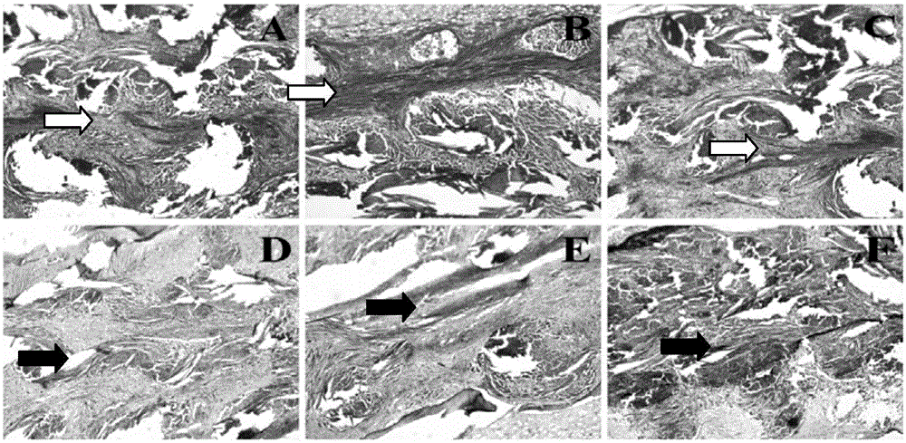
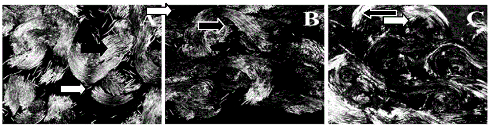
[0018] Preparation and transplantation of silk ligament grafts: knitting silk threads with flat needles on a knitting machine to form a network structure, after desericin, freeze-drying silk solution in partitions to create a spongy structure (inner area), and modifying hydroxyapatite after the silk solution is freeze-dried stone (external zone). After irradiation and disinfection, bone marrow mesenchymal stem cells were planted in the inner area, osteoblasts were planted in the outer area, and then rolled into a cylindrical shape and implanted into the established rabbit anterior cruciate ligament defect model.

[0019] 1) Samples were collected 1, 2, and 3 months after the implantation. When the samples were collected, the animals were sacrificed, and about 5 cm of the distal end of the femur and the proximal end of the tibia after the transplantati...


PUM


Abstract

The invention provides a method for staining hard tissue slices using picrosirius red and application of method. The method includes the following steps: sampling from a silk tissue engineering ligament, fixing and coating the sample using resin, performing hard tissue slicing, detecting the secretion condition of types I and III collagen through picrosirius red staining. According to the invention, the picrosirius red is used to stain the hard tissue slices, so that the sample processing time is shortened, the operation steps are simplified, and the types I and III collagen can be directly observed on the hard tissue slices.

Description

technical field [0001] The invention belongs to the field of biotechnology, and in particular relates to a method for detecting type I and type III collagens of silk tissue engineering ligaments. Background technique [0002] The anterior cruciate ligament plays a very important role in the normal movement and stability of the knee joint. The ability to heal after an ACL injury is minimal and often requires graft reconstruction surgery. The sources of ligament grafts mainly include autologous or allogeneic ligament grafts and tissue engineered ligaments. Commonly used sources of autologous ligament grafts are insufficient, and allogeneic ligament grafts are prone to immune rejection. Silk tissue engineered ligaments have attracted more and more attention due to their good mechanical properties, degradability and low immune rejection. [0003] After the silk ligament graft is implanted in the body, it is often necessary to detect the production of type I and III collagen t...

Claims


Application Information

Patent Timeline
Patent Type & Authority Applications(China)
IPC IPC(8): G01N1/30
Inventor 范宏斌孙立国李宏国屈玲郭征薛英森刘鑫成李征宇
Owner FOURTH MILITARY MEDICAL UNIVERSITY
Why Eureka
Try Eureka