
Multiplexed analyte detection

a technology of analyte detection and complexed analyte, which is applied in the direction of material analysis, biochemistry apparatus and processes, fermentation, etc., can solve the problems of low throughput, cumbersome assay of nucleic acids, and generally less stable assay conditions, so as to improve the dynamic range, improve the sensitivity to conventional methods, and improve the effect of resistance to contamination

Inactive Publication Date: 2005-04-14
AMPLIFIED PROTEOMICS
View PDF13 Cites 109 Cited by
  • Summary
  • Abstract
  • Description
  • Claims
  • Application Information

AI Technical Summary

Benefits of technology

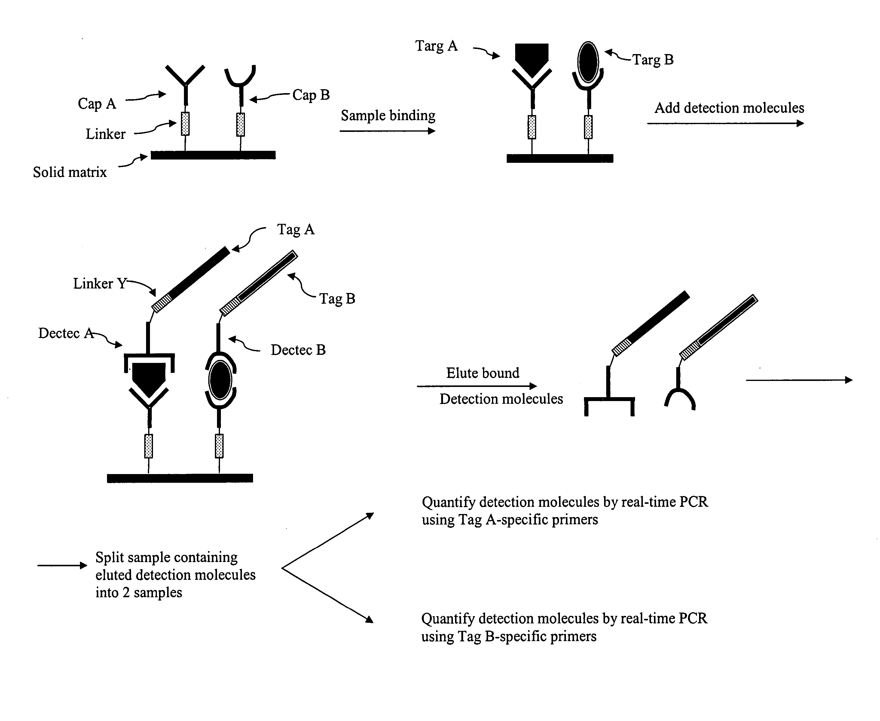
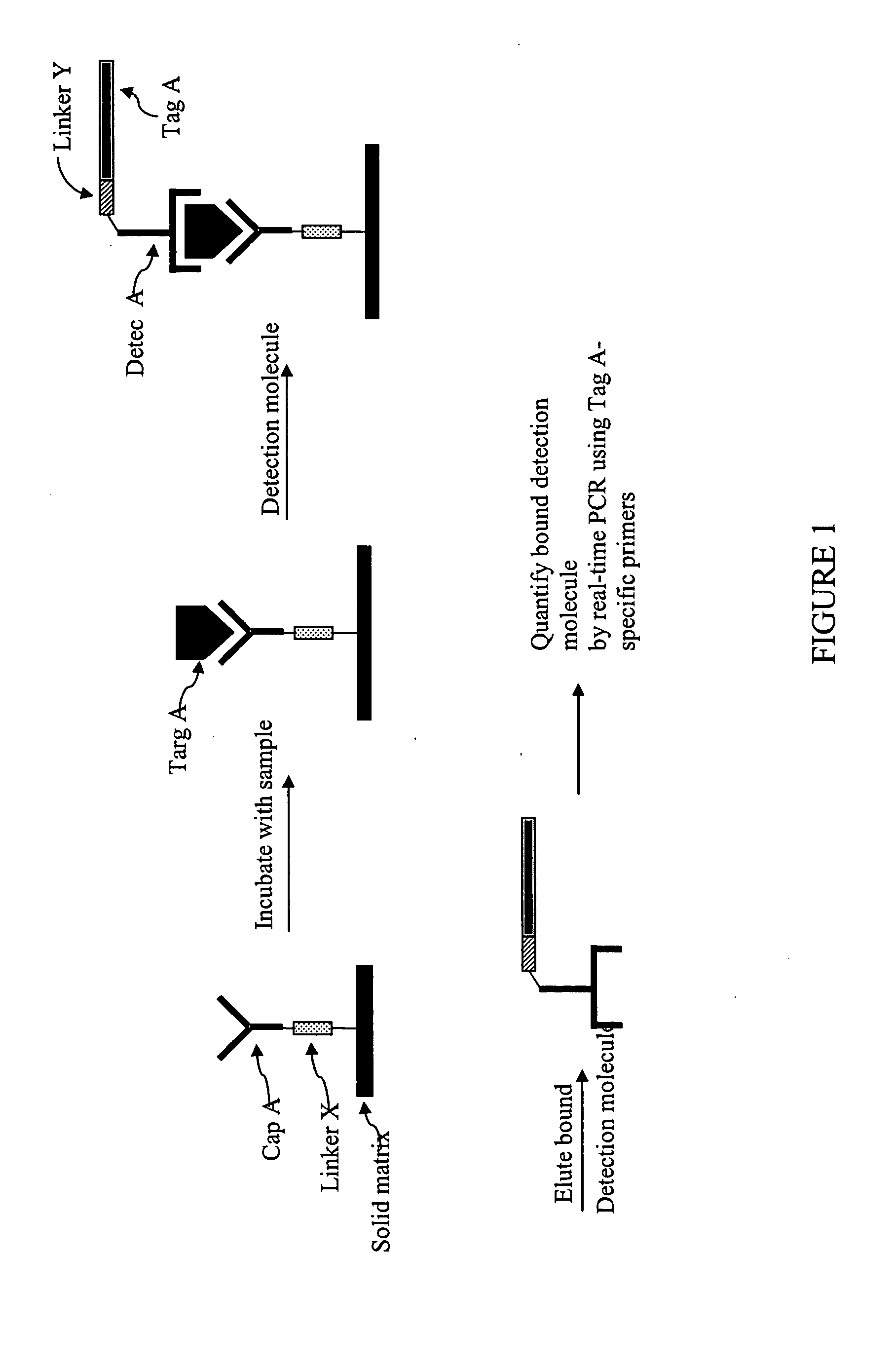
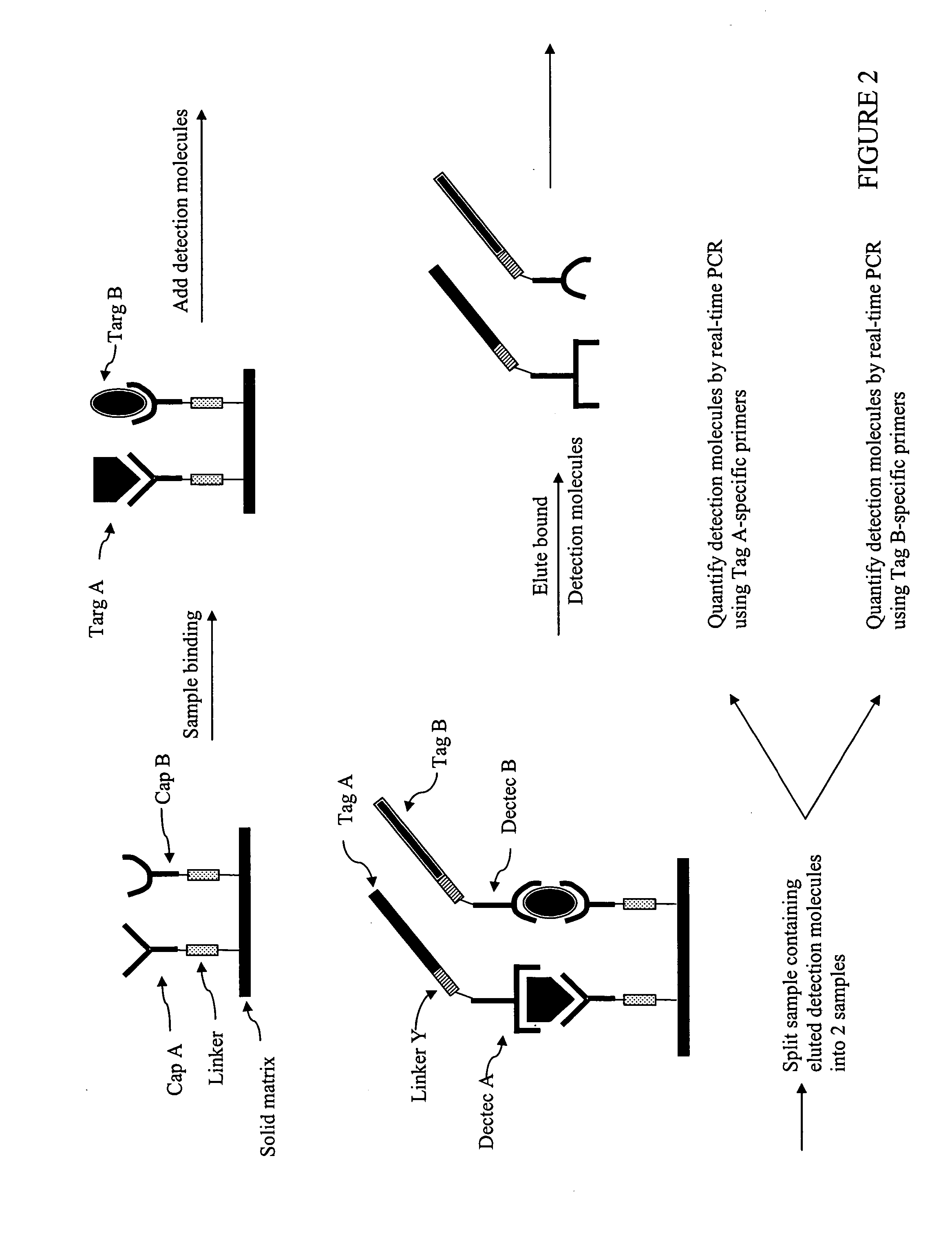
[0049] Accordingly, one object of the present invention is to provide a quantitative method for detecting the amounts of a plurality of different target molecules in a sample where the method has equal or improved sensitivity to conventional methods, has improved dynamic range, has improved resistance to contamination, has improved detection and which requires fewer human manipulation steps.
[0065] Alternatively, by using multicolor probes in real time PCR reactions, it is possible to detect 2, 3, 4 or more different nucleic acid sequence tags in a single PCR reaction containing 2, 3, 4 or more different primer sets for the sequence tags and using probes with different sequences, respectively. This form of multiplexing would reduce the total number of real time PCR reactions that would need to be run in order to measure the n different target molecules.
[0075] The methods of the invention are improvements over other multiplexed immunoassay platforms for the following reasons: the dynamic range and sensitivity are higher due to the immense amplification potential of nucleic acids; there is no need to prepare microscopic features derivatized with protein capture molecules, a process that is very difficult to achieve reproducibly; the equipment and procedures used are very similar to those performed in many laboratories and does not require unusual equipment or training.

Problems solved by technology

Nucleic acids are generally less stable under assay conditions and are not found readily in free form in body fluids.
Assays for nucleic acids have been cumbersome, with low through-put, poor specificity and poor quantitative ability.
One disadvantage of the antibody capture assay as described above is that the target molecules in the sample must be immobilized onto a solid support.
Although assays using antibodies are very useful, it is generally accepted that the detection limit of an assay is limited by the Kd of the antibody used as the capture molecule (Griswold, W.
As the concentration of analyte decreases to this sensitivity limit, the low percentage of capture molecules with bound analyte is insufficient to produce a detectable signal to noise ratio.
Conventional PCR amplification is not a quantitative detection method, however.
These impurities must be separated, usually with gel separation techniques, from the amplified product resulting in possible losses of material.
Although methods are known in which the PCR product is measured in the log phase (Kellogg et al., 1990; Pang et al, 1990), these methods require that each sample have equal input amounts of nucleic acid and that each sample amplifies with identical efficiency, and are therefore, not suitable for routine sample analyses.
However, this method could not be applied to biological samples due to the absence of a specific analyte capture molecule.
The addition of the five assay reagents plus washing, PCR amplification and detection resulted in an assay that was laborious and was subjected to both stoichiometric and disassociation complications (Hendrickson et al., 1995).
The direct linkage of the amplicon decreased the reagents used in the assay and the stoichiometric and disassociation complications that can occur.
However, these methods still require significant post-PCR manipulations, adding to increased labor and the very real possibility of laboratory contamination.
While immuno-PCR has provided sensitivities exceeding those of conventional ELISA, purification of the amplified product by gel electrophoresis requires substantial human manipulation and is, therefore, time-consuming.
Quantitation of the DNA label by analyzing the endpoint PCR product is prone to errors since the rate of product formation decreases after several cycles of logarithmic growth (Ferre, 1992; Raeymakers et al., 1995) and the post PCR sample handling may lead to laboratory contamination.
In the context of a immuno-PCR analysis, all of these amplicon quantitation techniques require significant post PCR analysis and induce the possibility of PCR product contamination of the laboratory for following assays because of the handling requirements.
Furthermore, these techniques are only able to analyze end-point PCR, PCR that has been stopped at a fixed PCR cycle number (e.g. 25 cycles of PCR).
This poses a problem in the dynamic range of amplicon quantitation because only some PCR reactions may be in the log phase of amplification; reactions with high amounts of template will have used all PCR reagents and stopped accumulating amplicon exponentially and reactions with low amounts of template might not have accumulated enough amplicon to be detectable.
Therefore, this phenomenon limits the detection range of PCR and can limit the analyte detection range of immuno-PCR assays.
The nuclease degradation of the hybridization probe releases the quenching of the reporter dye resulting in an increase in peak emission from the reporter.
An unfortunate problem in some selections is the maturation of non-specific ligands, or ligands that bind to the nitrocellulose filter or other material in the selection procedure.
It is clear that miss primed PCR artifacts will accumulate in the selection pool that is subjected to such a large amount of PCR cycling.
Labeling of the oligonucleotide with a reporter enzyme, however, requires additional chemical synthesis steps and additional labor, difficulties also associated with assays which use antibody reagents as described above.
Simple PCR amplification of a nucleic acid ligand provides additional quantities of the ligand, but has the disadvantage of requiring further separation steps to distinguish between the amplified ligand of interest and amplified nucleic acid impurities and primer dimers.
Traditional gel separation requires intensive manual labor.
Further, replicate experiments are required for statistical analysis and require additional time and labor.
These problems exist for both DNA ligands and RNA ligands used in these oligonucleotide assays.
The use of labeled primers allows detection of the PCR product, but does not overcome the problems of impurity and primer dimer amplification and is, therefore, not quantitative.
The main limitation to the method of Dodge is that it measures a single analyte molecule.
All of the abovementioned systems for the parallel quantification of multiple proteins suffer from limitations.
For the 2D arrays, difficulties arise in the preparation of reproducible microspots of capture reagent.
Also, the sensitivity of the 2D arrays or the bead-based systems is limited since it is not generally possible to use amplification methods for detection.
In particular, it is not possible to take advantage of the almost limitless amplification ability of nucleic acids.
Finally, none of the systems mentioned above can measure the presence of cell surface markers with a large dynamic range.
Of the three configurations mentioned above, only the homogenous solution-based methods can accurately quantitate cell surface marker amounts, but it is limited in terms of sensitivity and dynamic range.

Method used


Image

  • Multiplexed analyte detection
  • Multiplexed analyte detection
  • Multiplexed analyte detection

Examples


example

Quantitation and Detection of Interleukin 5, Interleukin 6, and TNF-α Using Sandwich-type Immunoassay and Real-time PCR

[0169] Primer, Probe, and DNA Labels

[0170] DNA labels and primers were synthesized by using standard phosphoramidite chemistry. An amino group was incorporated at the 5′ end of the DNA labels through a C6 spacer during the DNA synthesis. To introduce a sulfhydryl through the reaction with the 5′ amino group of the DNA labels, the DNA labels were dissolved in PBS buffer (10 mM sodium phosphate, 150 mM NaCl, pH 7.4) and Sulfo-LC-SPDP (sulfosuccinimidyl 6-[3′-(2-pyridyldithio)-propionamido]hexanoate (Pierce Endogen, Rockford, Ill.) was added to the DNA solutions to a final concentration of 2 mM. The reactions were carried out at room temperature for 1 hour. The 2-pyridylthio group protecting the sulfhydryl was subsequently removed by 5.5 mM TCEP (Tris[2-carboxyethylphosphine]hydrochloride) (Pierce Endogen, Rockford, Ill.) at room temperature for 1 hour. The DNA label...


PUM

PropertyMeasurementUnit
molecular weightaaaaaaaaaa
temperaturesaaaaaaaaaa
concentrationsaaaaaaaaaa

Abstract

The invention provides methods and kits for quantitating or detecting the presence of a plurality of different target molecules in a sample using detector molecules comprising nucleic acid tags.

Description

CROSS-REFERENCE TO RELATED APPLICATIONS [0001] This application claims the benefit of U.S. provisional application Ser. No. 60 / 489,213, filed Jul. 21, 2003, which is incorporated in its entirety by reference.FIELD OF THE INVENTION [0002] The invention relates to a method for detecting the presence of multiple target molecules in a sample which may contain the target molecules, using nucleic acid-containing detector molecules, amplification and quantitation or detection of the detector molecules. BACKGROUND OF THE INVENTION [0003] Numerous assays are known for detecting analytes in a sample. Two general types of analytes are protein analytes and nucleic acid analytes. The technology and assays directed at detecting proteins have historically developed separately and largely independently from the technology and assays directed at detecting nucleic acids. Several reasons exist for this trend in these two fields. Initially, proteins and nucleic acids are chemically distinct and have ve...

Claims


Application Information

Patent Timeline
Patent Type & Authority Applications(United States)
IPC IPC(8): C12P19/34C12Q1/68G01N
CPCC12Q1/6804C12Q1/6834C12Q2563/179C12Q2531/113C12Q2565/501C12Q2565/549C12Q2561/113
Inventor WU, JIE
Owner AMPLIFIED PROTEOMICS
Learn More