Preparation method of glucan microsphere gel
A technology of glucan and spherogel, applied in the field of preparation of hydrogel microspheres, can solve the problems of excessive hydrolysis of PVAc, unfavorable industrial production, difficult process control, etc., and achieves stable properties, narrow particle size distribution, Controllable range of effects
- Summary
- Abstract
- Description
- Claims
- Application Information
AI Technical Summary
Problems solved by technology
Method used
Image
Examples
Embodiment 1
[0057] This embodiment includes a synthesis technique of cross-linked dextran microsphere gel with specific pore size, including the following steps:
[0058] 1) Prepare 2.5M sodium hydroxide solution: Weigh 10g of sodium hydroxide, add 100ml of distilled water, and stir until colorless and transparent. Weigh 6.0±0.5g into a clean container;
[0059] 2) Prepare the dispersed phase: Weigh 3.0±0.3g of dextran, add it to the 2.5M sodium hydroxide solution, and stir under the homogenizing emulsification equipment. The stirring parameter is 600±100rpm, 3-5min. The sugar is completely dissolved into a colorless and transparent solution, which is used as the dispersed phase for use.
[0060] 3) Weigh 60±5g castor oil and place it in a clean container for use.
[0061] 4) Preparation of continuous phase: Weigh out 802.5±0.3g of Span, add it to the castor oil mentioned above, stir under homogeneous emulsification equipment, the stirring parameter is 600±100rpm, 3-5min, at this time Span 80 is...
Embodiment 2
[0071] The dextran microsphere gel of this example is basically the same as that of example 1, except that the concentration of the dispersed phase is changed: in this example, the mass of dextran added in step 2) is 6.0±0.6 g.
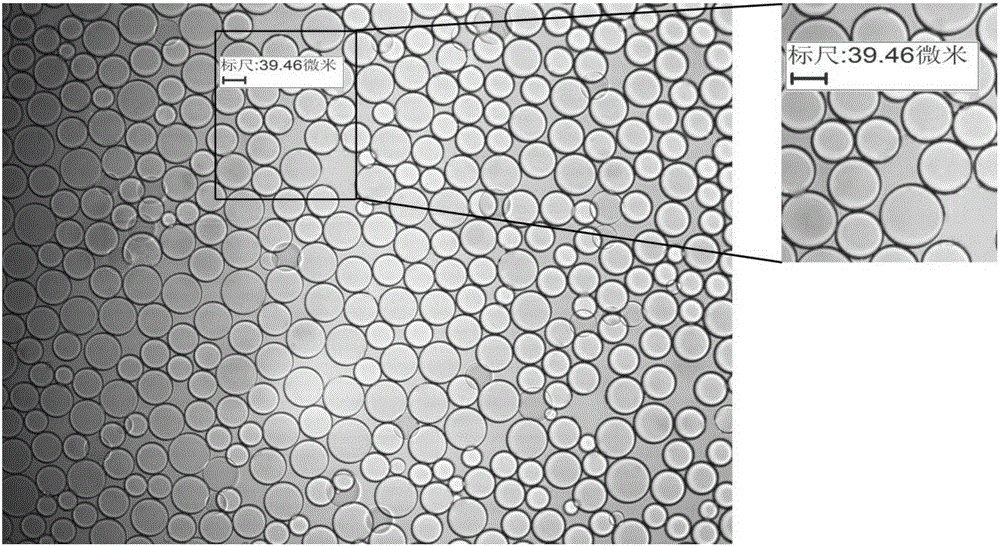
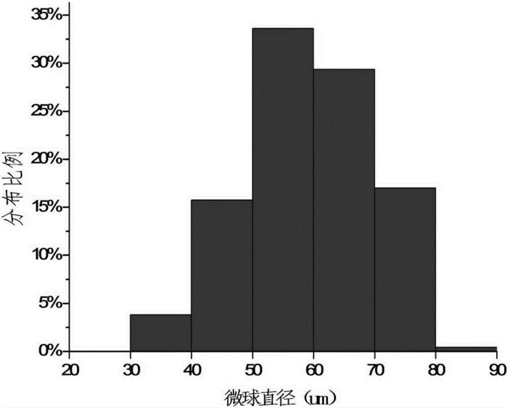
[0072] Observe the morphology of the dextran microspheres prepared by the method of this embodiment with a biological microscope at 100 times, such as Figure 4 , Randomly select three microspheres for measurement, and get their radii respectively 39.62μm, 23.08μm, 48.40μm; take a sample to measure the particle size range of the gel microspheres, and the particle size distribution range is as follows Figure 5 ,, 40-120μm, can be applied to serological detection in the field of clinical diagnosis, such as microcolumn gel method to detect specific size infectious disease markers, cancer syndrome markers, etc., and for proteins, polysaccharides, and nucleic acids of different molecular weights , Separation and purification of enzymes and other substances.
Embodiment 3
[0074] The dextran microsphere gel of this example is basically the same as that of Example 1, and the difference is that the dispersing and emulsifying speed is changed: in this example, step 6) adjusts the homogeneous emulsification equipment parameter to 600±100rpm, 25- 30min.
[0075] Observe the morphology of the dextran microspheres prepared by the method of this embodiment with a biological microscope at 100 times, such as Image 6 , Randomly select three microspheres for measurement, and get their radii respectively 23.60μm, 33.94μm, 49.81μm. Sampling to measure the particle size range of the gel microspheres, the particle size distribution range is as follows Figure 7 , 40-120μm, can be used for serological testing in the field of clinical diagnosis, such as micro-column gel method to detect specific size infectious disease markers, cancer syndrome markers, etc., and used for proteins, polysaccharides, nucleic acids, Separation and purification of enzymes and other subst...
PUM
| Property | Measurement | Unit |
|---|---|---|
| Radius | aaaaa | aaaaa |
| Radius | aaaaa | aaaaa |
Abstract
Description
Claims
Application Information
 - R&D
- Intellectual Property
- Life Sciences
- Materials
- Tech Scout
- Unparalleled Data Quality
- Higher Quality Content
- 60% Fewer Hallucinations
Browse by: Latest US Patents, China's latest patents, Technical Efficacy Thesaurus, Application Domain, Technology Topic, Popular Technical Reports.
© 2025 PatSnap. All rights reserved.Legal|Privacy policy|Modern Slavery Act Transparency Statement|Sitemap|About US| Contact US: help@patsnap.com


