

Breast cancer cell dyeing method, application thereof and dyeing kit
A technique for breast cancer cells and a staining method, which is applied in the field of breast cancer cell staining methods and applications and staining kits, can solve problems such as poor prognosis, and achieve the effects of good application prospects, simple operation and good accuracy.
- Summary
- Abstract
- Description
- Claims
- Application Information
AI Technical Summary
Problems solved by technology
Method used
Image

Examples
Embodiment 1
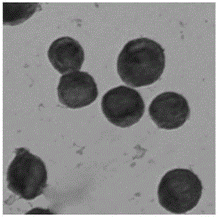
[0048] Example 1 Staining of breast cancer cell lines.
[0049] (1) First prepare a glass slide on which the breast cancer cell line MCF-7 is tiled on the surface: digest the breast cancer cell line MCF-7 cultured in vitro, add 1 ml of PBS solution, dilute and mix well.
[0050] (2) Turn on the Hettich centrifuge, add samples with the PBS containing breast cancer cell line MCF-7 in step (1), so that the total cell volume of each sample well is 0.3M, and discard the PBS solution after centrifugation to obtain a surface The slides of breast cancer cell MCF-7 were tiled, air-dried, and fixed with acetone for 10 min.
[0051] (3) Use an immunohistochemical pen to draw a circle around the tissue of the slide obtained in step (2), the circle is about 3mm away from the tissue, wash with PBS for 3min, and then wash with 3wt%H 2 o 2 Soak at room temperature for 10 minutes, and then wash with PBS 3 times, 3 minutes each time, to obtain a glass slide with breast cancer cells spread on ...
Embodiment 2
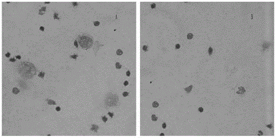
[0056] Example 2 Staining detection of breast cancer cell line MCF-7 added to normal peripheral blood samples.
[0057] 1) Collect 5ml of peripheral blood sample with a sodium heparin anticoagulant tube, add a small amount of PBS solution containing breast cancer cell line MCF-7, and mix well.
[0058] 2) Centrifuge in a centrifuge (3500 rpm, 5 minutes), absorb and discard the upper layer of plasma, and obtain about 2-3ml of blood cells.
[0059] 3) After diluting and mixing the remaining blood cell components with 3ml PBS, add 15ml Ficoll (lymphocyte separation medium, Lymphoprep, Axis-Shield PoCAS, Oslo, Norway) into a centrifuge tube, and centrifuge for 20min in a horizontal centrifuge (22°C, 2000 rpm, 0 speed to start, 0 speed to decelerate to stop).
[0060] 4) Take out the specimen from the centrifuge, extract the mononuclear cell layer cells, add to the PBS solution and mix well, centrifuge (2000 rpm, 10min), discard the original PBS, and add 2ml of new PBS to dilute a...
Embodiment 3
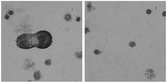
[0063] Example 3 Staining detection by adding breast cancer cell lines to bone marrow blood samples.
[0064] 1) Collect 5ml of bone marrow blood sample with a sodium heparin anticoagulant tube, add a small amount of PBS solution containing breast cancer cell line MCF-7, and mix well.
[0065] 2) Centrifuge in a centrifuge (3500 rpm, 5 minutes), absorb and discard the upper layer of plasma, and obtain about 2-3ml of blood cells.
[0066] 3) After diluting and mixing the remaining blood cell components with 3ml PBS, add 15ml Ficoll (lymphocyte separation medium, Lymphoprep, Axis-Shield PoCAS, Oslo, Norway) into a centrifuge tube, and centrifuge for 20min in a horizontal centrifuge (22°C, 2000 rpm, 0 speed to start, 0 speed to decelerate to stop).
[0067] 4) Take out the specimen from the centrifuge, extract the mononuclear cell layer cells, add to the PBS solution to mix, centrifuge (2000 rpm, 10min), discard the original PBS, add 2ml of new PBS to dilute and mix, spare.
...
PUM
 Abstract
Description
Claims
Application Information