Real-time ultrasonic elasticity imaging method and system

An ultrasonic elastography and image technology, applied in the directions of ultrasonic/sonic/infrasonic Permian technology, ultrasonic/sonic/infrasound image/data processing, organ motion/change detection, etc., can solve the relationship between displacement and image gradient, Poor approximation results, discontinuous image displacement, etc.

Active Publication Date: 2016-01-27
CHISON MEDICAL TECH CO LTD
View PDF8 Cites 11 Cited by
  • Summary
  • Abstract
  • Description
  • Claims
  • Application Information

AI Technical Summary

Problems solved by technology

If these two assumptions are not true, that is, the image is discontinuous and the displacement is large, the displa

Method used


Image

  • Real-time ultrasonic elasticity imaging method and system
  • Real-time ultrasonic elasticity imaging method and system
  • Real-time ultrasonic elasticity imaging method and system

Examples


Embodiment Construction

[0066] The present invention will be further described below in conjunction with specific drawings and embodiments.

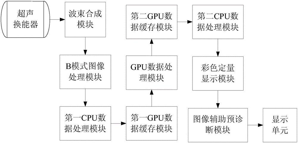
[0067] The real-time ultrasonic elastography method proposed by the present invention is jointly realized by CPU and multi-core GPU (GraphicProcessingUnit), and the method steps are as follows:

[0068] S10. Ultrasonic scanning of the target area of ​​the biological tissue and receiving echo signals when the biological tissue is slowly squeezed by the ultrasonic transducer;

[0069] S20. Perform A / D conversion, in-phase superposition, and other processing on the received echo electrical signal in step S10 through the beamforming module to form line data;

[0070] S30. Perform noise reduction, filtering, and signal-to-noise ratio processing on the line data obtained in step S20 through the B-mode image processing module to form B-mode images in different states of deformation of biological tissues after being slowly squeezed by an external force;

[0071] S40. ...


PUM


Abstract

The invention provides a real-time ultrasonic elasticity imaging method and system. The imaging method mainly comprises the steps of obtaining B-mode images under different states of deformation of biological tissues caused by slow extrusion of external force; selecting ROI areas of two frame images; obtaining displacement fields by using the optical flow method and transmitting parameter information of sub-steps requiring acceleration operations and containing repetitive operations to a first GPU data caching module in the calculation process; performing the operation treatment of all the sub-steps requiring acceleration operations with a GPU working group; putting the operation result information of all the sub-steps into an optical flow method operation process and finally obtaining the displacement fields, namely, the optical flow fields, of the ROI areas; obtaining the axial strain fields of the ROI areas of the images; performing noise reduction treatment on axial strain field information; performing colorizing treatment on obtained information; performing elastic classification on target areas in the ROI areas. The real-time ultrasonic elasticity imaging method has the advantages of high operation speed, relatively high precision and relatively great robustness.

Description

technical field [0001] The invention relates to the field of ultrasonic echo imaging, in particular to a real-time ultrasonic elastic imaging method and system. Background technique [0002] Ultrasonic echo imaging technology has been widely used in military, medical and other fields. The concept of elastography in ultrasound echo imaging was first proposed by Ophir et al. in 1991. After that, elastography technology has developed rapidly in the past two decades, and it is called the E-mode after the A-mode, B-mode, D-mode, and M-mode ultrasound. Ultrasound elastography is the imaging of biological tissue elasticity parameters through an ultrasonic imaging system. Ultrasound elastography can provide biological tissue elastic characteristics that cannot be reflected by traditional B-ultrasound images, and is very helpful for clinical applications such as tumor detection. Due to the advantages of easy implementation, suitable for real-time diagnosis, and non-invasiveness to ...

Claims


Application Information

Patent Timeline
IPC IPC(8): A61B8/08
Inventor 孙新赵明昌
Owner CHISON MEDICAL TECH CO LTD
Why Eureka
Try Eureka