Preparation and preservation method of canine and amniotic membrane

The technology of canine amniotic membrane and amniotic membrane is applied in the field of preparation and preservation of canine amniotic membrane, which can solve the problems of ineffective preparation effect and good canine amniotic membrane, and achieve the effects of good biocompatibility and long effective usable time.

Inactive Publication Date: 2016-04-27
金艺鹏
View PDF3 Cites 5 Cited by
  • Summary
  • Abstract
  • Description
  • Claims
  • Application Information

AI Technical Summary

Problems solved by technology

[0004] What the present invention aims to solve is the technical proble...

Method used


Image

  • Preparation and preservation method of canine and amniotic membrane

Examples


Embodiment 1

[0017] Embodiment 1, a preparation method of canine amniotic membrane, the aseptically obtained canine amniotic membrane is placed in sterile physiological saline solution;

[0018] Repeated washing with a large amount of sterile 0.9% sodium chloride saline solution to wash away excess blood, mucus and chorionic tissue fragments on the amniotic membrane;

[0019] Use sterilized phosphate buffer solution PBS whose main components are Na2HPO4, KH2PO4, NaCl and KCl to wash repeatedly to wash away visible blood vessels and blood clots. After washing, the amniotic membrane should be uniform in color and soft and translucent.

[0020] The following steps are also included: placing the washed amniotic membrane in 1% polyethylene glycol octylphenyl ether TritonX-100 solution to seal, and placing it in a constant temperature shaker at 37°C for 24 hours;

[0021] Rinse thoroughly with phosphate-buffered PBS solution whose main components are Na2HPO4, KH2PO4, NaCl and KCl, add 0.25% tryp...

Embodiment 2

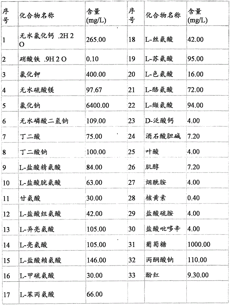
[0033] Embodiment 2, a method for preserving canine amniotic membrane, the present invention also includes a method for preserving canine amniotic membrane, mixing various amino acids and glucose DMEM medium with pure glycerol 1:1, and using a 20 micron pore size filter membrane to filter Obtain about 4 milliliters of sterile preservation solution and divide into sterile 5ml centrifuge tubes, put the canine amnion prepared according to the above claim 1 or 2 into the centrifuge tubes with preservation solution respectively, so that the preservation solution is completely infiltrated Canine amnion, sealed and stored in a -80°C refrigerator.

[0034] Quality Inspection of Preserved Canine Amniotic Membrane

[0035] Take out the freshly prepared canine amniotic membranes after storage for 1 month, 3 months, and 6 months, rewarm them at room temperature for 10 minutes, wrap them on coverslips, and put 0.25% pentadiene After being fixed in aldehyde solution, samples were prepared ...


PUM


Abstract

The invention discloses a preparation and preservation method of a canine and amniotic membrane, and relates to the technical field of a tissue engineering material, aiming at solving the technical problem of the prior art which cannot prepare a canine and amniotic membrane which is good in effect. The technical scheme comprises the following steps: keeping a canine and amniotic membrane which is obtained in a sterile mode in a sterile physiological saline solution; repeatedly flushing with membrane with a great amount of sterile 0.9% sodium chloride physiological saline solution, so that residual blood stains, mucus, chorion tissue fragments on the amniotic membrane are flushed away; and repeatedly flushing the membrane by virtue of a sterilized phosphate buffered solution PBS which contains Na2HPO4, KH2PO4, NaCl and KCl as main ingredients, so that visible blood vessels and blood clots are cleaned, wherein the cleaned amniotic membrane is uniform in color and is in the form of a soft semi-transparent membrane.

Description

technical field [0001] The invention relates to the technical field of tissue engineering materials, in particular to a preparation and preservation method of canine amniotic membrane. Background technique [0002] The amniotic membrane is the innermost layer of the placenta, smooth and elastic, with a thickness of about 0.02 to 0.5 mm. The amniotic membrane can be divided into five layers: a single layer of columnar epithelial cells, a dense basement membrane layer, an acellular layer rich in collagen, and a layer rich in collagen types I, III, IV, V, and VII, and laminin. connective tissue layer. The connective tissue layer is divided into three parts: an acellular layer with a dense network of fibers; a loose network of fibroblasts with sporadically distributed fibroblasts; and a spongy layer. These include the chorion as well as complex fibers and are wrapped in mucus. Some animal experiments and clinical observations have shown that the basement membrane of human amn...

Claims


Application Information

Patent Timeline
IPC IPC(8): A61L27/36A01N1/02
Inventor 金艺鹏白鹤王亚楠
Owner 金艺鹏
Why Eureka
Try Eureka