
Method for measuring single alive myocardial cell action potential and pulsing force by atomic force microscope

An atomic force microscope and cardiomyocyte technology, applied in the field of engineering, can solve the problems of difficulty in locating electrodes and cells, and inability to measure the electrical properties of cells, and achieve the effect of stable sealing impedance

Active Publication Date: 2016-04-27
CHANGCHUN UNIV OF SCI & TECH
View PDF5 Cites 16 Cited by
  • Summary
  • Abstract
  • Description
  • Claims
  • Application Information

AI Technical Summary

Problems solved by technology

However, the position of the electrodes in the microelectrode array is fixed, and the cells grow randomly on the electrodes, so it is difficult to locate the positions of the electrodes and the cells, so that the electrical characteristics of the selected cell positions cannot be measured

Method used


Image

  • Method for measuring single alive myocardial cell action potential and pulsing force by atomic force microscope
  • Method for measuring single alive myocardial cell action potential and pulsing force by atomic force microscope
  • Method for measuring single alive myocardial cell action potential and pulsing force by atomic force microscope

Examples


Embodiment Construction

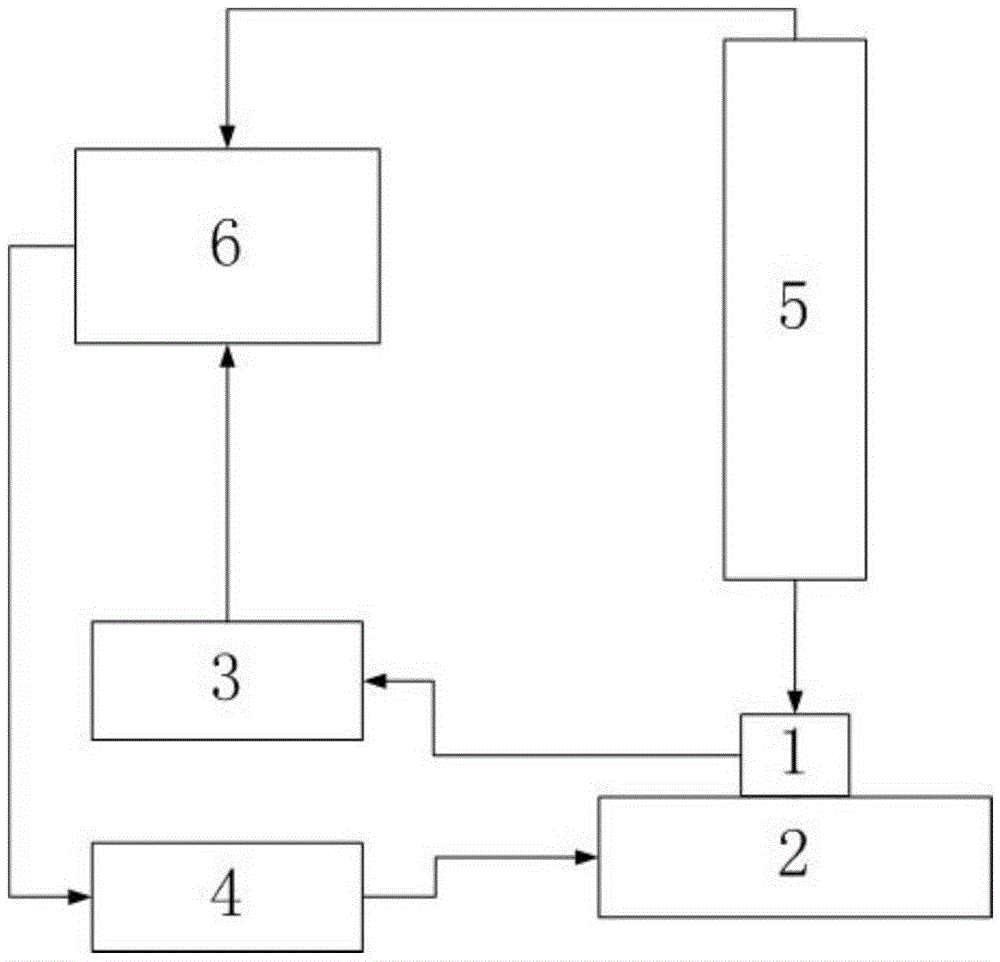
[0031] Such as figure 1 As shown, it is a block diagram of the principle of the present invention, wherein 1 is a conductive probe measuring cell module, including an optical lever system composed of an atomic force microscope conductive probe, cardiomyocytes, lasers and four-quadrant photodetectors, and 2 is a piezoelectric ceramic nano-displacement platform , 3 is a four-quadrant photoelectric detector voltage acquisition and myocardial cell action potential acquisition module, 4 is a piezoelectric ceramic nano-displacement platform control module, 5 is an optical microscope module, and 6 is a computer control system;

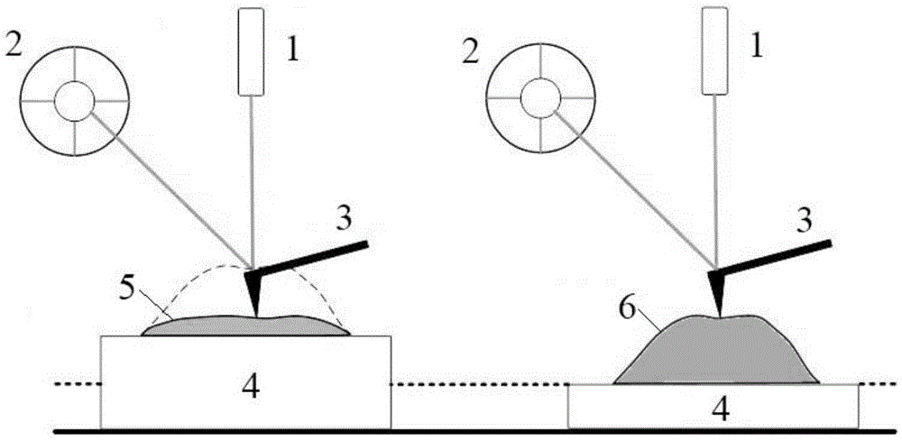
[0032] Such as figure 2 As shown, it is a schematic diagram of the present invention's atomic force microscope conductive probe tracking cardiomyocyte beating, wherein 1 is a laser, 2 is a four-quadrant photodetector, 3 is an atomic force microscope conductive probe, 4 is a sample stage, and 5 is a diastolic state 6 is the cardiomyocyte in the contraction s...


PUM


Abstract

The invention relates to the method for measuring single alive myocardial cell action potential and the pulsing force by an atomic force microscope. An atomic force microscope conductive probe is used as a nanometer electrode to measure the operation potential of the single pulsing mouse myocardial cell; the probe is accurately positioned on a cell to be detected through the atomic force microscope which is independently developed and according to a force feedback system; the probe and the cell surface have two contact forms which are a constant force contact mode and a non-constant force contact mode; and the variable measurement of the action potential and the pulsing force is performed on the myocardial cell through the two modes. The measurement on the myocardial cell action potential can accurately determine the position of the myocardial cell, and realizes the fixed point measurement. The invention can obtain the high signal-noise ratio action potential signal waveform and can simultaneously measure the action potential of the myocardial cell, ie, the changing signal of the pulsing force.

Description

technical field [0001] The invention relates to a method for measuring the action potential and beating force of a single live cardiomyocyte with an atomic force microscope, which belongs to the technical field of engineering. Background technique [0002] With the development of nanotechnology, atomic force microscopy (AFM), as an important tool for nanoscale measurement, makes its application in biomedicine possible, especially in the research on the manipulation and characteristic detection of living cells, which shows that it is relatively Advantages unique to other live cell characterization techniques. [0003] The electrophysiological properties of cells are the properties of charged cells such as cardiomyocytes and nerve cells. Measuring the electrophysiological properties of such cells can be used for the diagnosis of heart and nerve diseases, regeneration and rehabilitation of cardiac muscle and nerve cells, and high-throughput drug screening. etc. can bring major...

Claims


Application Information

Patent Timeline
Patent Type & Authority Applications(China)
IPC IPC(8): G01Q60/24G01Q60/40
CPCG01Q60/24G01Q60/40
Inventor 王作斌张思微王国梁王馨悦曲英敏赵立张楠张志坚董莉彤宋正勋翁占坤
Owner CHANGCHUN UNIV OF SCI & TECH
Learn More