Opto-acoustic quantitative elasticity imaging method and device

A technology of elastography and photoacoustics, which is applied in the field of biomedical detection, can solve the problems of not being able to obtain the elastic modulus and reduce the accuracy of measurement results, and achieve the effect of ensuring high-sensitivity detection, high accuracy, and high detection sensitivity

Active Publication Date: 2016-05-11
SOUTH CHINA NORMAL UNIVERSITY
View PDF5 Cites 12 Cited by
  • Summary
  • Abstract
  • Description
  • Claims
  • Application Information

AI Technical Summary

Problems solved by technology

However, in the above method, the quantitative measurement of the elastic modulus of the tissue cannot be realized, that is, the absolute value of the elastic m...

Method used


Image

  • Opto-acoustic quantitative elasticity imaging method and device
  • Opto-acoustic quantitative elasticity imaging method and device
  • Opto-acoustic quantitative elasticity imaging method and device

Examples


Embodiment 1

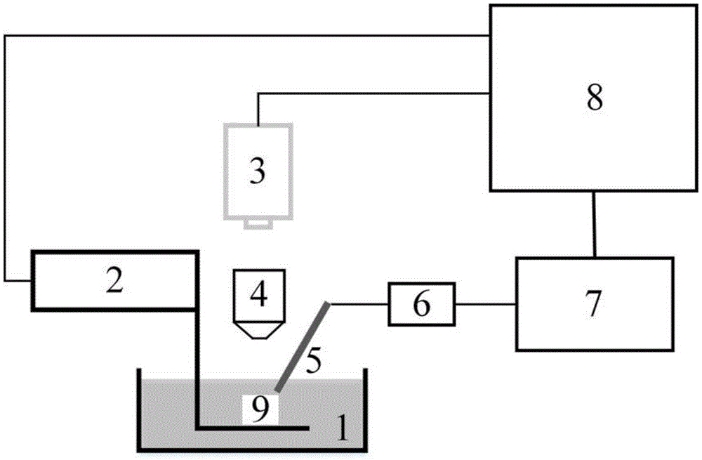
[0043] Such as figure 1 As shown, the photoacoustic quantitative elastography device of this embodiment includes a photoacoustic excitation source, a signal acquisition / transmission / reconstruction assembly, a coupling tank 1, a stepper motor 2, an X-Y two-dimensional scanning platform and an instrument fixing / supporting instrument assembly (Fig. not shown), the photoacoustic excitation source includes a laser 3 and a focusing lens 4; the signal acquisition / transmission / reconstruction assembly includes an ultrasonic probe 5, an amplifier 6, an oscilloscope 7 and a computer 8, and the ultrasonic probe 5 , amplifier 6, oscilloscope 7 and computer 8 are connected successively; The sampling rate of described oscilloscope 7 is 2.5GHz; Described computer 8 is equipped with acquisition control and signal processing system, and this system utilizes Labview and Matlab programming to form; Described step The motor 2 is connected to the computer 8, the X-Y two-dimensional scanning...

Embodiment 2

[0067] The present embodiment is the experiment that utilizes agar sample to carry out, mainly comprises the following steps:

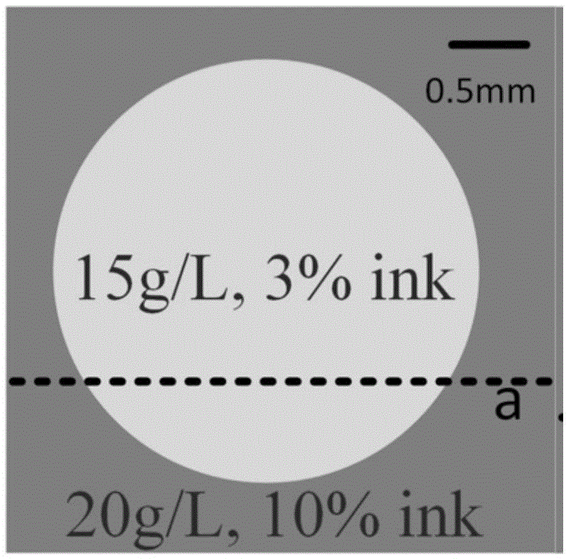
[0068] 1) Add 10% ink to make a square sample in the agar with a concentration of 20g / L, make agar with a concentration of 15g / L in the middle of it and add 3% ink to make a square sample, which forms image 3 Agar sample a in the agar; Adding 10% ink to make a square sample in the agar with a concentration of 30g / L, making agar with a concentration of 25g / L in the middle of it and adding 3% ink to make a square sample, this formed Figure 4 Agar sample b in agar;

[0069] 2) start the laser, the output pulse laser has a wavelength of 532nm, a pulse width of 10ns, and a repetition rate of 15Hz; the pulse laser is irradiated on agar samples a and b after being focused by a focusing lens, and agar samples a and b are excited to emit photoacoustic signals, The photoacoustic signal is received by the ultrasonic detector after passing through the...


PUM

PropertyMeasurementUnit
Diameteraaaaaaaaaa
Thicknessaaaaaaaaaa
Sampling rateaaaaaaaaaa

Abstract

The invention discloses an opto-acoustic quantitative elasticity imaging method and device. The method comprises the following steps: a laser device emits pulse laser which is focused by a focusing lens and irradiates a tissue sample so as to excite an opto-acoustic signal, and the opto-acoustic signal is received by an ultrasonic detector through a coupling solution in a coupling trough; the opto-acoustic received by the ultrasonic detector is collected by an oscilloscope after being amplified by an amplifier, and signal information is stored into a computer; the computer controls a stepping motor to move the tissue sample point by point, so that an X-Y two-dimensional planar scanning region is formed on the corresponding tissue sample; after the oscilloscope collects all the signals, the computer calculates a quantitative elastic modulus of the tissue sample of each point; and a quantitative elastic two-dimensional image of the tissue sample is reconstructed according to the calculated quantitative elastic modulus. The device comprises an opto-acoustic exciting source, a signal collection/transmission/reconstruction assembly, the coupling trough, the stepping motor and an X-Y two-dimensional scanning platform. The opto-acoustic quantitative elasticity imaging method and device can realize non-destructive and high-resolution elastic and quantitative tissue measurement and imaging.

Description

technical field [0001] The invention relates to an elastography method and device, in particular to a photoacoustic quantitative elastography method and device, belonging to the technical field of biomedical detection. Background technique [0002] The pathological changes of tissues are often accompanied by the changes of their mechanical properties, especially the elastic modulus, so the detection of tissue lesions can be realized by analyzing the physical parameters of tissues. At present, the method of predicting tissue diseases by detecting tissue elasticity has been widely accepted by doctors and imaging researchers. For example, the detection method of non-destructive liver cirrhosis using ultrasound elasticity detection method has been applied to the parameter detection of clinical diseases. [0003] The existing quantitative elastic testing methods are mainly static ultrasonic elastic testing and ultrasonic shear wave elastography. The static ultrasonic elastic det...

Claims


Application Information

Patent Timeline
IPC IPC(8): G01N21/17
CPCG01N21/1702G01N2021/1706G01N2021/1708
Inventor 杨思华杨芬石玉娇邢达
Owner SOUTH CHINA NORMAL UNIVERSITY
Why Eureka
Try Eureka