A minimally invasive intervention method for establishing a heart failure animal model

A minimally invasive intervention and animal model technology, applied in the field of biomedicine, can solve problems such as the difficulty of suturing and fixing pacemaker electrodes, the animal survival rate of less than 60%, and the difficulty of thoracotomy surgery, so as to facilitate physical recovery , easy anti-infection treatment, beneficial to healing effect

Inactive Publication Date: 2016-06-22
SICHUAN KANGCHENG BIOLOGICAL SCI & TECH
View PDF0 Cites 7 Cited by
  • Summary
  • Abstract
  • Description
  • Claims
  • Application Information

AI Technical Summary

Problems solved by technology

The common thoracotomy methods for installing a cardiac pacemaker are sternal median surgery and left thoracic wall surgery. These two thoracotomy operations have great risks, involving harsh anesthesia conditions, difficult thoracotomy techniques, and suture fixation. The...

Method used


Image

  • A minimally invasive intervention method for establishing a heart failure animal model
  • A minimally invasive intervention method for establishing a heart failure animal model
  • A minimally invasive intervention method for establishing a heart failure animal model

Examples


Embodiment 1

[0036] Example 1: Minimally invasive intervention method for installing canine pacemaker

[0037] 1. Anesthesia

[0038] Inject 3% sodium pentobarbital solution intravenously, 1mL / kg, that is, 30mg / kg, observe the breathing of the experimental dogs.

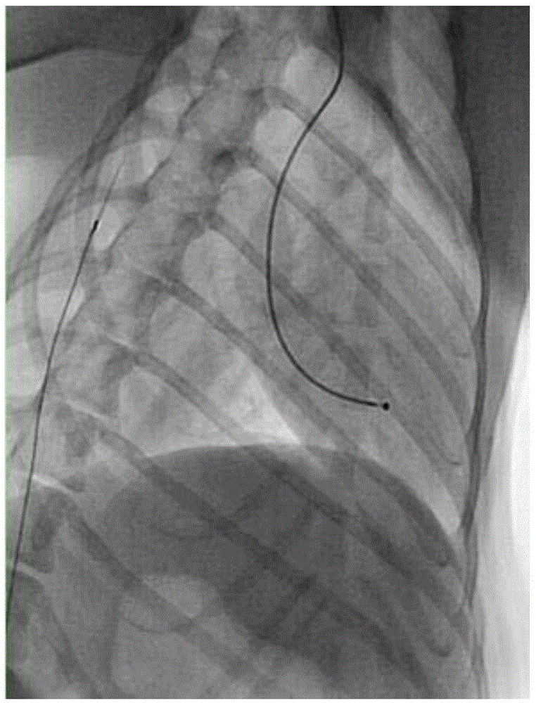
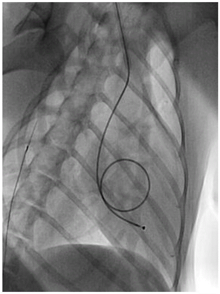
[0039] 2. Install pacemaker electrodes

[0040] The dog was placed in a lying position on the left side, and the right neck was shaved, 75% alcohol was used for skin preparation, and iodophor disinfection. Use a scalpel to make an incision parallel to the skin about 2 cm parallel to the body, and use gauze to stop bleeding. The right common jugular vein was bluntly separated, the common jugular vein was punctured with a vascular puncture needle, and a 5F catheter sheath was inserted to establish a vascular cannula. The distal end of the right common jugular vein was completely ligated with sutures, and the blood vessel wall of the proximal right common jugular vein was slightly tightened and fixed with the catheter sheath with sutures....

Embodiment 2

[0045] Example 2: Establishment of a model of canine heart failure

[0046] 1. Color Doppler Doppler Inspection Results

[0047] Pacing time

End diastolic volume EDV (mL)

End-systolic volume ESV (mL)

Stroke volume per minute SV (mL)

Left ventricular fractional shortening FS (%)

Ejection fraction EF (%)

Before pacing

19

6

14

38

71

Pacing for 1 week

45

16

29

34

64

Pacing for 2 weeks

84

46

38

22

46

Pacing for 3 weeks

56

33

23

20

41

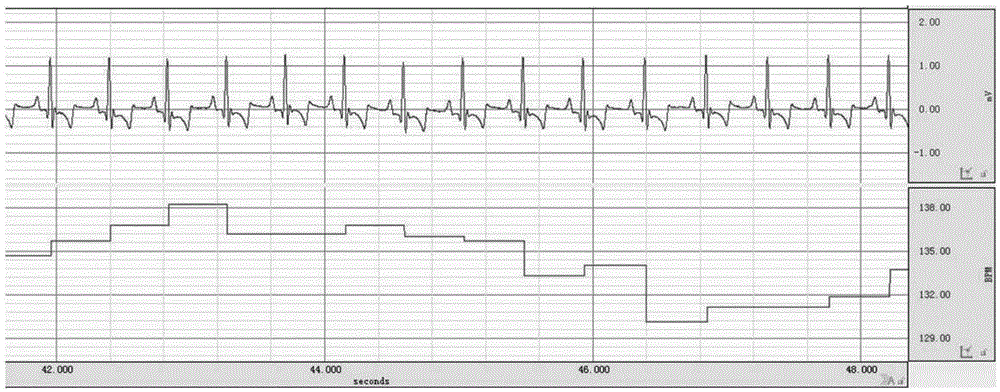
[0048] 2. ECG examination results of canine heart failure model

[0049] Install a pacemaker to establish a dog heart failure model. ECG before pacing ( image 3 ), ECG after 1 week of pacing ( Figure 4 ), ECG after 2 weeks of pacing ( Figure 5 ), ECG after 3 weeks of pacing ( Image 6 ).

[0050] 3. Myocardial enzyme test results of canine heart failure model

[0051] Pacing time

ALT (U / L)

AST (U / L)

CK (U / L)

CKMB (U / L)

LDH (U / L)

UERA (mmol / L)

CREA (μmol / L)

TBIL (μmol / L)

Before pacing

27.75

34.85

358.80

349.70

106.60

2.92

37...


PUM


Abstract

Animal models of heart failure are established, which are often used for preclinical drug efficacy evaluation and mechanism research of heart failure therapeutic drugs. The present invention provides a simple and reliable minimally invasive intervention method for establishing an animal heart failure model. By implementing the minimally invasive intervention method, the animal is installed with a cardiac pacemaker, which avoids excessive trauma to the experimental animal during the thoracotomy operation, and reduces the The complexity of pacemaker installation and postoperative care costs have greatly improved the survival rate of animals after pacemaker installation, thereby ensuring the success rate of establishing animal models of heart failure.

Description

Technical field [0001] The invention belongs to the field of biomedicine, and particularly relates to a minimally invasive intervention method for establishing an animal model of heart failure. Background technique [0002] Heart failure (heart failure) is the final stage of clinical development of various cardiovascular diseases. The annual fatality rate of this disease is about 5%~10%, which seriously threatens human health. The establishment of a heart failure animal model by a simple and reliable method is very beneficial to the preclinical evaluation of the efficacy of the treatment of heart failure and the study of the mechanism of action. At present, it is common to use surgical thoracotomy to implant a cardiac pacemaker and rapid right ventricular pacing to construct an animal model of heart failure. The installation method of a pacemaker is the key technology for establishing an animal model of heart failure. [0003] The conventional technical process of constructing can...

Claims


Application Information

Patent Timeline
IPC IPC(8): A61D1/00A61N1/372
CPCA61D1/00A61N1/362A61N1/372
Inventor 肖凯张印兵张琰艳王俊龙钟治晖叶雪飞王莉
Owner SICHUAN KANGCHENG BIOLOGICAL SCI & TECH
Why Eureka
Try Eureka