Biliary tract exploration method and device based on superfine endoscope

A bile duct and channel technology, applied in endoscopy, laparoscopy, medical science and other directions, can solve the problems of guide wire displacement, difficulty in operation, inability to operate, etc., and achieve the effect of preventing gastric coiling, reducing pain, and maintaining good stability.

Inactive Publication Date: 2016-09-21
刘时助
View PDF7 Cites 5 Cited by
  • Summary
  • Abstract
  • Description
  • Claims
  • Application Information

AI Technical Summary

Problems solved by technology

According to literature search, domestic and foreign scholars in the prior art use ultra-fine endoscopes to enter the common bile duct. There are mainly the following methods: 1. Guide wire guidance—Gastrointest Endosc.2006 May; Endoscopic retrograde cholangiopancreatography (encoscopicretrograde cholangio-pancreatography, ERCP) was performed with papillary sphincterotomy or columnar balloon dilatation, and the duodenoscope was withdrawn after dilation to 10-12 mm. Duodenoscope retains the guide wire, and under the guidance of X-rays, inserts an ultra-fine endoscope along the guide wire and enters the common bile duct through the duodenal papilla. In this technique, the disadvantage of guide wire displacement is common
2. Anchoring balloon guidance—Gastrointest Endosc.2009 Aug;70(2):297-302, insert a guide wire or a 5 Fr detachable balloon catheter into the intrahepatic bile duct or In the upstream of the stenosis, inflate the balloon to anchor the balloon above the intrahepatic bile duct or the stenosis, then separate the handle at the proximal end of the balloon catheter from the catheter and properly block the catheter to prevent balloon leakage, exit ten Duodenoscope and guide wire, and then insert the endoscope into the bile duct along the balloon catheter in a "J" shape. During the intervention, the balloon needs to be withdrawn from the working channel, which will cause the position of the endoscope in the bile duct to be incorrect. Stable. In addition, anchoring the balloon in the bile duct is difficult in some cases, especially in those with bile duct stricture or marked dilation of the common bile duct; this method has been discontinued due to complications of air embolism and biliary rupture ;3. Outer cannula assistance—Endoscopy.2012 Nov.44(11):1037-44, a side hole is opened at a distance of 65-75 cm from the distal end of the outer cannula to allow the endoscope to pass through, and the endoscope is inserted into the overcoat through the side hole Insert the tube together with the outer cannula into the duodenal bulb. After inflating and anchoring the outer cannula balloon, the gastroscope is withdrawn, and the ultra-fine endoscope is inserted into the duodenum with sphincterotomy or balloon expansion The main nipple. However, compared with ultra-thin endoscopes, the available overtubes are very large, and it is difficult to operate through the overtubes
The repeatability of this operation method needs to be further verified, and it is difficult to continue to explore the microscope
[0005] In summary, the above operation methods have their own advantages and disadvantages, and cannot make the operation simple and stable, and meet the needs of repeated access to the biliary tract for diagnosis and treatment.

Method used


Image

  • Biliary tract exploration method and device based on superfine endoscope
  • Biliary tract exploration method and device based on superfine endoscope
  • Biliary tract exploration method and device based on superfine endoscope

Examples


Embodiment 2

[0046] Embodiment 2, a method of biliary tract exploration based on an ultra-fine endoscope in this embodiment is suitable for cases where it is difficult to guide a guide wire into the common bile duct with an ultra-fine endoscope, and the steps are as follows:

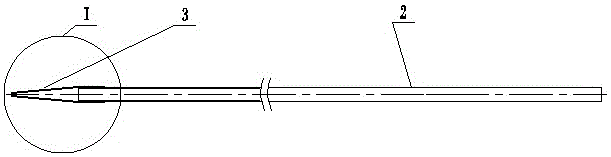
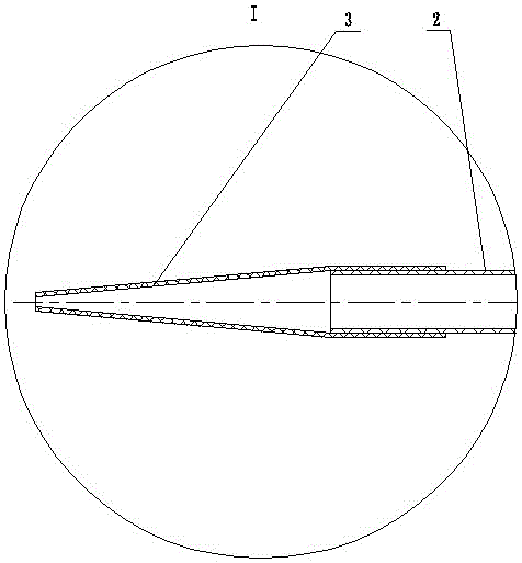
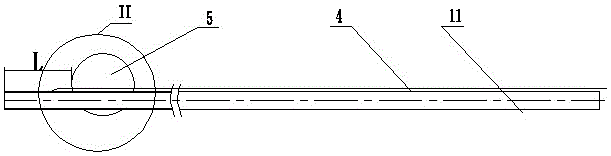
[0047] 1) After incision and dilation of the duodenal papilla by routine ERCP of the duodenoscope, the front end of the ultra-fine endoscope 2 is fixed to the transparent tapered guide head 3, and then the ultra-fine endoscope 2 is put on the outer tube within 4;

[0048] 2) Ultra-thin endoscope 2 with transparent tapered guide head 3 and sheathed with outer tube 4 enters the descending part of the duodenum through the mouth, pharynx, esophagus, and stomach 8, and passes through the duodenal papilla. Play J-shaped reversal;

[0049] 3) When performing a J-shaped reversal, use the traction wire 11 to pull back the overtube 4, and after the ultra-fine endoscope 2 finds the opening of the duodenal papilla, the transpar...


PUM


Abstract

The invention provides a biliary tract exploration method and device based on a superfine endoscope. The biliary tract exploration method is characterized by comprising the following steps that a conical guide head is fixedly connected, and the superfine endoscope is mounted in an inserted mode; the superfine endoscope is put into the duodenum descending part; the conical guide head is fixed to the open position of duodenal papilla; a guide wire is inserted, and the superfine endoscope is brought into the common bile duct and the common hepatic duct by the guide wire; an annular surrounding saccule is opened through inflation, and an outer sleeve is fixed; the guide wire and the superfine endoscope are taken out and the outer sleeve is dwelled to form a stable biliary tract exploration channel. The biliary tract exploration device comprises the guide wire, a duodenoscope and the superfine endoscope and is characterized by further comprising the conical guide head, wherein the conical guide head is arranged at the front end of the superfine endoscope and is fixedly connected with the superfine endoscope, the annular surrounding saccule and a traction wire are arranged at the front portion of the outer sleeve, the superfine endoscope and the guide wire are inserted into the outer sleeve and enter the common bile duct, and the annular surrounding saccule is opened through inflation to fix the outer sleeve so as to form the biliary tract exploration channel.

Description

technical field [0001] The invention relates to a diagnosis and treatment method and a medical device, and is a biliary tract exploration method and a device thereof based on an ultrafine endoscope. Background technique [0002] At present, POC-peroral cholangioscopy can be divided into three categories: ①Peroral cholangioscopy. ② spyglass choledochoscopy. ③ ultra-fine endoscopy. Compared with the above two kinds of choledochoscope and traditional angiography, the ultra-fine endoscope entering the biliary tract has the following advantages: 1. In terms of diagnosis: large field of view, clear images, water injection and air injection, easy to observe; 2. In terms of treatment: high-definition Directly looking at laser lithotripsy, early biliary tract lesions can be treated endoscopically, and common bile duct tumors can be clearly observed and biopsied, guiding the guide wire through the narrow biliary tract, and assisting in the placement of biliary stents; 3. The equipme...

Claims


Application Information

Patent Timeline
Patent Type & Authority Applications(China)
IPC IPC(8): A61B1/313A61B1/00A61M25/09
CPCA61B1/3132A61B1/00131A61B1/00154A61M25/09
Inventor 刘时助王宏光陈宁金世柱张训兵王曼彤郭庆梅谢延侠汪强郭享孙震刘俊鹏夏润辉
Owner 刘时助
Why Eureka
Try Eureka