

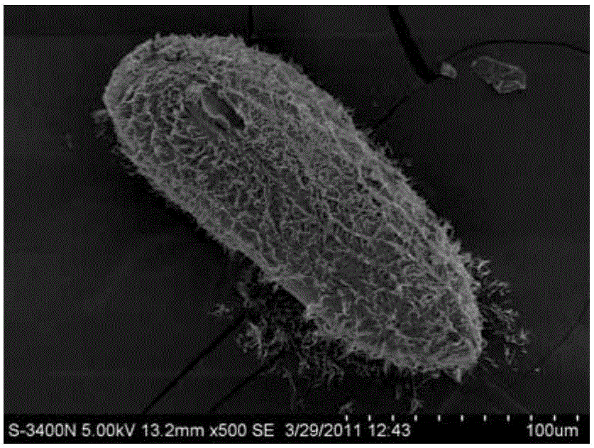
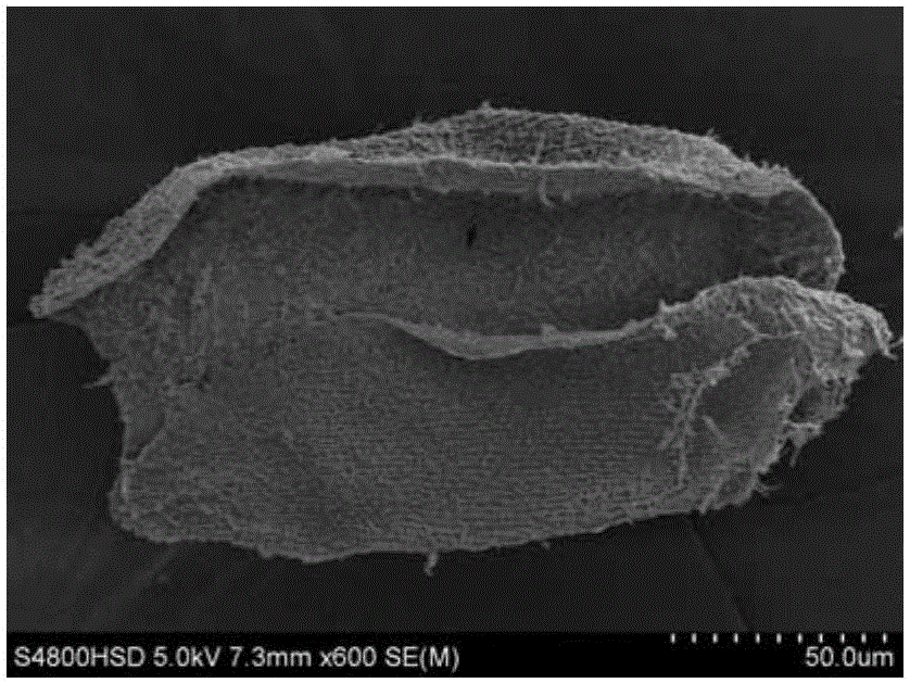
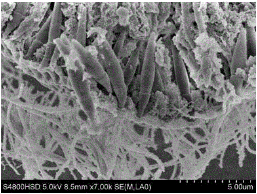
Method for membrane breaking production of protozoa single cell scanning electron microscopy sample
A technology of protozoa and scanning electron microscopy, which is applied in the direction of measuring devices, instruments, scientific instruments, etc., can solve the problems of internal structure damage of cells, loss of organelles, and inability to observe the structure of cytoplasm, etc., and achieve good in-situ characteristics and simple dehydration steps , the effect of reducing the experimental risk
- Summary
- Abstract
- Description
- Claims
- Application Information
AI Technical Summary
Problems solved by technology
Method used
Image

Examples
specific Embodiment approach 1
[0057] Embodiment 1: The method for preparing a protozoan single-cell scanning electron microscope sample by breaking the membrane in this embodiment is carried out according to the following steps:
[0058] 1. Material collection and cleaning
[0059] The day before the experiment, the protozoa were treated with low temperature or starvation, and on the second day, 140-160 protozoa with normal morphology and uniform protoplasm were picked into a clean concave dish, and the protozoa bodies were rinsed with pure water to remove the protozoa. Clean the dirt attached to the surface of the body, and then suck out the water in the concave dish, leaving as little water as possible;
[0060] 2. Fixation and membrane rupture
[0061] Inject the fixed membrane-breaking agent into the fixed cylinder, use a micropipette to absorb the protozoa in the concave dish and inject it into the fixed cylinder, fix it in the refrigerator at 4°C in the dark and rupture the membrane for 10-15 minute...
specific Embodiment approach 2
[0074] Embodiment 2: The difference between this embodiment and Embodiment 1 is that the low temperature treatment in Step 1 is at 16-18° C. for 24 hours. Others are the same as in the first embodiment.
specific Embodiment approach 3
[0075] Embodiment 3: This embodiment is different from Embodiment 1 in that: the starvation treatment in Step 1 is to stop feeding for 24-48 hours. Others are the same as in the first embodiment.
PUM
| Property | Measurement | Unit |
|---|---|---|
| Diameter | aaaaa | aaaaa |
| Height | aaaaa | aaaaa |
| Diameter | aaaaa | aaaaa |
Abstract
Description
Claims
Application Information