Application of hpse gene in screening pigs resistant to PRRS
A technology for PRRSV and PRRSV, which is applied in the field of biomedicine, can solve the problems of PRRSV prevention and control, the lack of cross-protection of vaccines, and damage to the immune system of the body, and achieve good clinical application value.
- Summary
- Abstract
- Description
- Claims
- Application Information
AI Technical Summary
Problems solved by technology
Method used
Image

Examples
Embodiment 1
[0040] Example 1 PRRSV infection inhibits the expression of heparan sulfate HS
[0041] 1. Cultivate Marc-145 cells with DMEM medium containing 10% fetal bovine serum to a confluence of 60-70%, wash once with PBS, replace with DMEM medium containing 2% fetal bovine serum, and set MOI=0.1 Add PRRSV at 37°C, 5% CO 2 Cultured in a humidified incubator for 0, 24, and 36 h. Wash 3 times with PBS, fix with 4% paraformaldehyde for 10 min, wash with PBS for 10 min, perforate with 10% Triton-100 for 15 min, wash with PBS for 10 min, then block with 1% BSA diluted in PBS for 30 min, add anti-HSPG2 protein for one Anti-mouse (diluted 1:200) was incubated overnight at 4°C, anti-mouse secondary antibody (diluted 1:1000) was used for 1 h, nuclei were stained with DAPI dye for 5 min, and washed with PBS for 10 min. First check whether the heparan sulfate HS is stained under an ordinary fluorescent inverted microscope, and then observe under a confocal microscope.
[0042] 2. The result is...
Embodiment 2
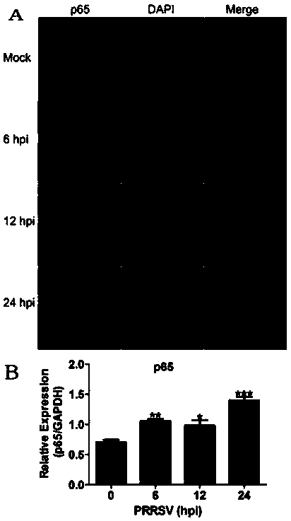
[0043] Example 2 PRRSV infection activates NF-κB signaling pathway
[0044] 1. Culture Marc-145 and PAMs cells with DMEM and RPMI 1640 medium containing 10% fetal bovine serum respectively to a confluence of 60-70%, wash once with PBS, and replace with DMEM and RPMI 1640 containing 2% fetal bovine serum Culture medium, and add PRRSV at a ratio of MOI=0.1 at 37°C, 5% CO 2 Cultured in a humidified incubator for 0, 6, 12, 24, and 36 h. The p65 subunit of the NF-κB signaling pathway was detected by confocal and qRT-PCR according to the above method, and the cells were collected for Western Blot detection of the IκBα subunit of the NF-κB signaling pathway.
[0045] 2. The result is as follows figure 2 with 3 As shown, after PRRSV infects PAMs cells, the p65 subunit of NF-κB signaling pathway enters the nucleus ( figure 2 A), increasing the expression of p65 subunit of NF-κB signaling pathway ( figure 2 B). After PRRSV infected PAMs and Marc-145 cells, the phosphorylation of...
Embodiment 3
[0047] Example 3 PRRSV infection promotes HPSE gene expression
[0048] 1. Culture Marc-145 and PAMs cells with DMEM and RPMI 1640 medium containing 10% fetal bovine serum respectively to a confluence of 60-70%, wash once with PBS, and replace with DMEM and RPMI 1640 containing 2% fetal bovine serum Culture medium, and add PRRSV at a ratio of MOI=0.1 at 37°C, 5% CO 2 Cultured in a humidified incubator for 0, 6, 12, 24, and 36 h. Cells were collected for Western Blot detection of heparanase encoded by HPSE gene.
[0049] 2. The result is as follows Figure 4 As shown, PRRSV infects PAMs ( Figure 4 A) and Marc-145 ( Figure 4 B) after cells, the expression level of heparanase Heparanase encoded by HPSE gene is gradually increased. That is, PRRSV promotes the expression of HPSE gene after infecting PAMs and Marc-145 cells.
[0050] The expression of HPSE gene increased, and the activity of heparanase increased, which in turn cut HS, inhibited PRRSV particles from adhering ...
PUM
 Abstract
Description
Claims
Application Information
 - R&D
- Intellectual Property
- Life Sciences
- Materials
- Tech Scout
- Unparalleled Data Quality
- Higher Quality Content
- 60% Fewer Hallucinations
Browse by: Latest US Patents, China's latest patents, Technical Efficacy Thesaurus, Application Domain, Technology Topic, Popular Technical Reports.
© 2025 PatSnap. All rights reserved.Legal|Privacy policy|Modern Slavery Act Transparency Statement|Sitemap|About US| Contact US: help@patsnap.com