Separation and detection methods of circulating tumor cells

A technology of tumor cells and separation methods, which is applied in the separation and detection of circulating tumor cells, does not depend on the field of blood cell tumor markers, can solve the problem of indetermination of cell invasion ability, etc., and achieves the effect of simple separation and detection methods

Inactive Publication Date: 2016-12-21
XIAMEN RENRUI BIOMEDICAL TECH CO LTD
View PDF3 Cites 9 Cited by
  • Summary
  • Abstract
  • Description
  • Claims
  • Application Information

AI Technical Summary

Problems solved by technology

However, this method is not based on actual migration or invasion, therefore

Method used


Image

  • Separation and detection methods of circulating tumor cells
  • Separation and detection methods of circulating tumor cells
  • Separation and detection methods of circulating tumor cells

Examples


Embodiment 1

[0044] Example 1 Detection of CTCs in mice transplanted with xenogeneic tumors

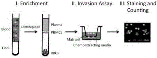
[0045]To develop a method for the isolation and detection of CTCs based on cell invasion ability, we first established a mouse model with metastatic tumors. A549, a non-small cell lung cancer cell line that stably expresses enhanced green fluorescent protein (GFP) and luciferase (Luc), was used to generate tumors in the flanks of nude mice. Surgical resection was performed when the flank tumors reached a diameter of 1 cm, and 60-70% of mice developed lung metastases 3 months after surgery.

[0046] Whole blood collection was performed 3 months after surgery. Whole blood was subjected to density gradient centrifugation using Ficoll lymphocyte separation medium to collect mononuclear cells (PBMCs), washed with serum-free RPMI-1640 cell culture medium, and PBMCs were resuspended.

[0047] The Transwell chamber invasion experiment was performed on the enriched PBMC: before the experiment, a layer of...

Embodiment 2

[0051] Example 2 Correlation between CTC detected by CICA and invasion and metastasis

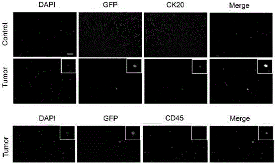
[0052] To demonstrate the utility of CICA, we tested CICA in a number of control and A549 xenografted nude mice. The whole blood of each transplanted tumor mouse and control mouse was collected, and CTC was detected by the method of CICA. We did not detect cytokeratin-positive CTCs in control mice. In contrast, blood samples from most tumor-implanted mice contained 22-78 CTCs positive for invasive GFP (Table 1). More interestingly, some mice transplanted with tumors had no detectable migrated CTCs (Table 1). These results indicated that CTCs isolated by CICA were associated with metastasis.

[0053] Table 1 CICA detection results of different mice

[0054]

Embodiment 3

[0055] Example 3 Comparison between CICA and EpCAM-based methods

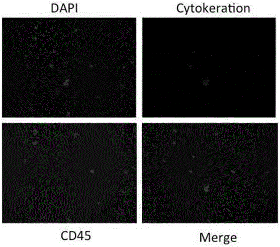
[0056] Currently, the most common isolation method for CTCs is immunomagnetic bead sorting using epithelial markers of CTCs. In order to compare these two methods, we divided the blood of mice transplanted with A549 tumor into two parts, and used these two methods to isolate CTCs respectively. The results showed that the EpCAM immunomagnetic bead method separated more than 40% of CTC cells than the CICA method (see Table 2).

[0057] However, when the CTCs screened by the immunomagnetic bead method were subjected to invasion experiments, only about 50% of the cells showed invasion ability. More interestingly, less invasive cells were screened by immunomagnetic beads than those isolated directly by CICA. Since immunomagnetic bead screening does not affect the invasive ability of cells, these results show that although more CTCs were obtained by anti-EpCAM antibody-based immune selection screening, many of them...


PUM


Abstract

The invention provides a separation method of circulating tumor cells (CTCs). The method comprises the following steps: enriching mononuclear cells (PBMC) from a blood sample through density gradient centrifugation; carrying out invasion experiments on the enriched PBMCs, and collecting invasive cells; and dyeing and sorting the collected cells. The invention also provides a detection method of the CTCs. The methods are not influenced by the heteroplasmy of the CTCs, the separated CTCs are tumor cells with invasion ability and are more than CTCs with invasion ability, obtained through an immunomagnetic bead screening method, the CTCs can be used in tumor transfer researches in the laboratory, and CTC detection can be used as the biomarker liquid biopsy of the cancer prognosis and treatment effect.

Description

technical field [0001] The invention belongs to the field of tumor research, and in particular relates to a new method for isolating and detecting circulating tumor cells (CTC), which is a method independent of blood cell tumor markers. Background technique [0002] Circulating tumor cells (circulating tumor cells, CTCs) refer to tumor cells that shed from the tumor and enter the circulation system. With the continuous proliferation of tumor cells, some cells can secrete a substance that inhibits the expression of adhesion factors, increase their motility and separate them from the tumor mother. These exfoliated tumor cells then secrete a proteolytic enzyme to destroy the peripheral hosts connective tissue and enters the vasculature. Diagnosis and treatment operations can also cause tumor cells to spread into the peripheral blood circulation. [0003] Recent studies suggest that CTCs contain cells capable of initiating the growth of metastatic tumors, the cause of death in...

Claims


Application Information

Patent Timeline
IPC IPC(8): C12N5/09G01N33/569
CPCC12N5/0693G01N33/56966
Inventor 许华曦卜国军
Owner XIAMEN RENRUI BIOMEDICAL TECH CO LTD
Why Eureka
Try Eureka