Preparation and testing method of a three-dimensional decompression device for avascular necrosis of the femoral head

A technology of decompression device and testing method, which is applied in the direction of bone drill guidance, medical science, surgery, etc., can solve the problems of increasing the amount of X-ray radiation of patients, prolonging the operation time, etc., and achieves the effect of easy promotion and reasonable production cost

Inactive Publication Date: 2019-01-15
THE AFFILIATED HOSPITAL OF SOUTHWEST MEDICAL UNIV
View PDF7 Cites 0 Cited by
  • Summary
  • Abstract
  • Description
  • Claims
  • Application Information

AI Technical Summary

Problems solved by technology

In the clinical practice of fine-needle porous decompression, we found that positioning each channel will greatly prolong the operation time, anesthesia time, and increase the patient's exposure to X-rays

Method used


Image

  • Preparation and testing method of a three-dimensional decompression device for avascular necrosis of the femoral head
  • Preparation and testing method of a three-dimensional decompression device for avascular necrosis of the femoral head
  • Preparation and testing method of a three-dimensional decompression device for avascular necrosis of the femoral head

Examples


preparation example Construction

[0027] see image 3 As shown, the present invention also provides a method for preparing a three-dimensional decompression device based on the above-mentioned avascular necrosis of the femoral head, comprising the following steps:

[0028] Step S101. Import the CT three-dimensional image data (general dicom format) of the patient into MIMICS software, extract the bone data of the proximal femur on the surgical side by the MIMICS software, and calculate and generate the 3D image of the proximal femur bone;

[0029] Step S102. Create a mask of the proximal femur bone in the MIMICS software, and set an expansion value of the mask; specifically, the expansion value of the mask is 6 mm.

[0030] Step S103. Apply the generated mask to subtract the original skeleton image to obtain a new mask for skeleton shelling; specifically, the thickness of the new mask for skeleton shelling is 6 mm.

[0031]Step S104. Use MIMICS software to edit the new mask of the bone shell, only keep the bo...

specific example

[0044] 1. Determination of the anatomical parameters of the normal human proximal femur

[0045] 100 cases of hip joints without deformity were randomly selected from the CT imaging system of our hospital. The proximal femoral neck shaft angle, femoral neck diameter, femoral head diameter, and femoral neck axis length were measured by the hospital CT imaging system to determine the data distribution interval of each parameter.

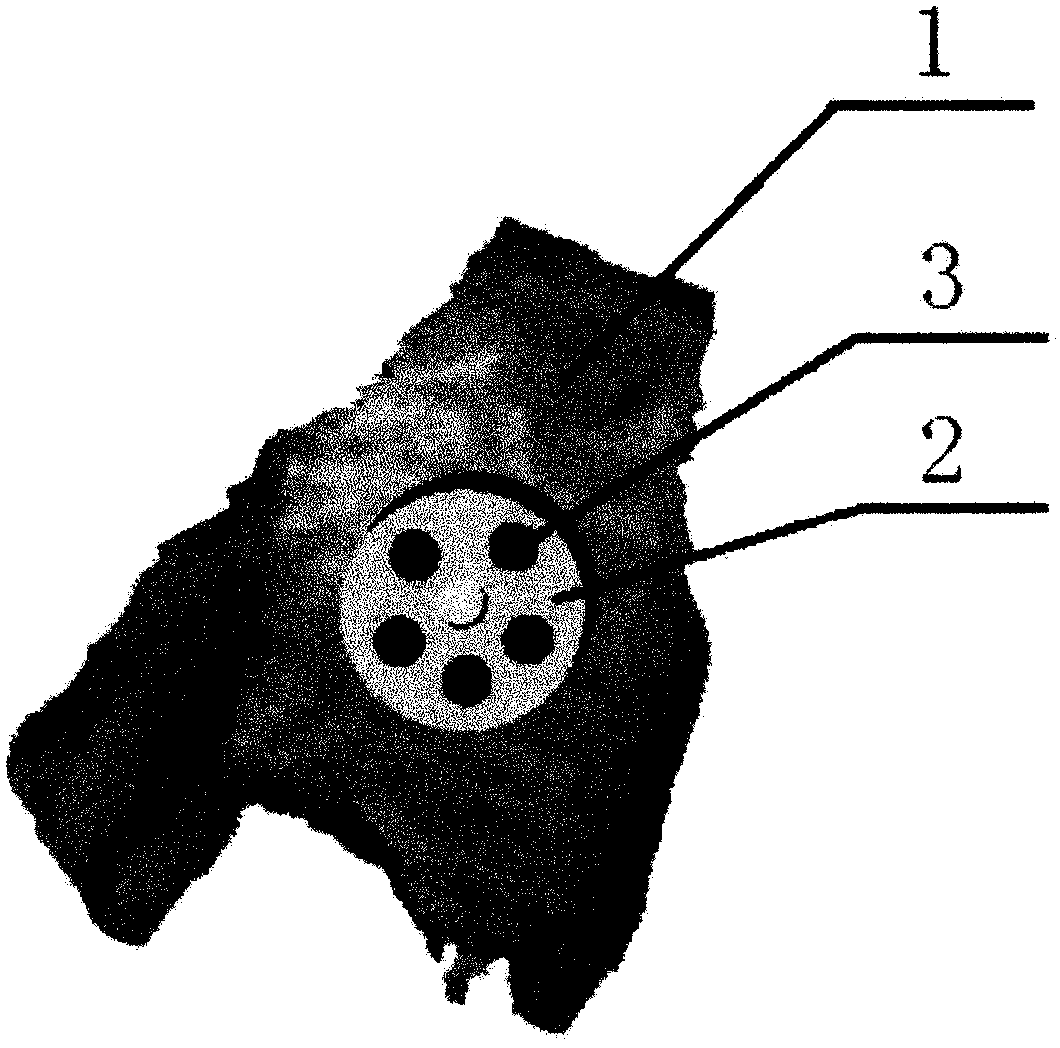
[0046] 2. Design guidance system and make guidance system

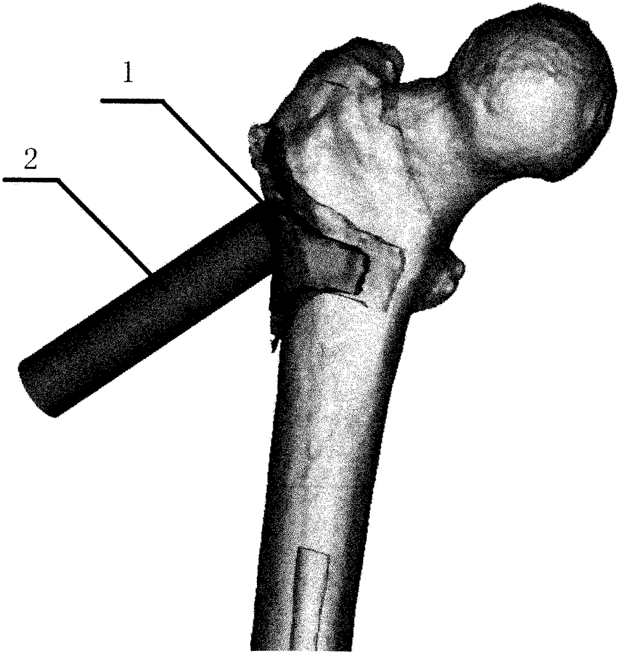
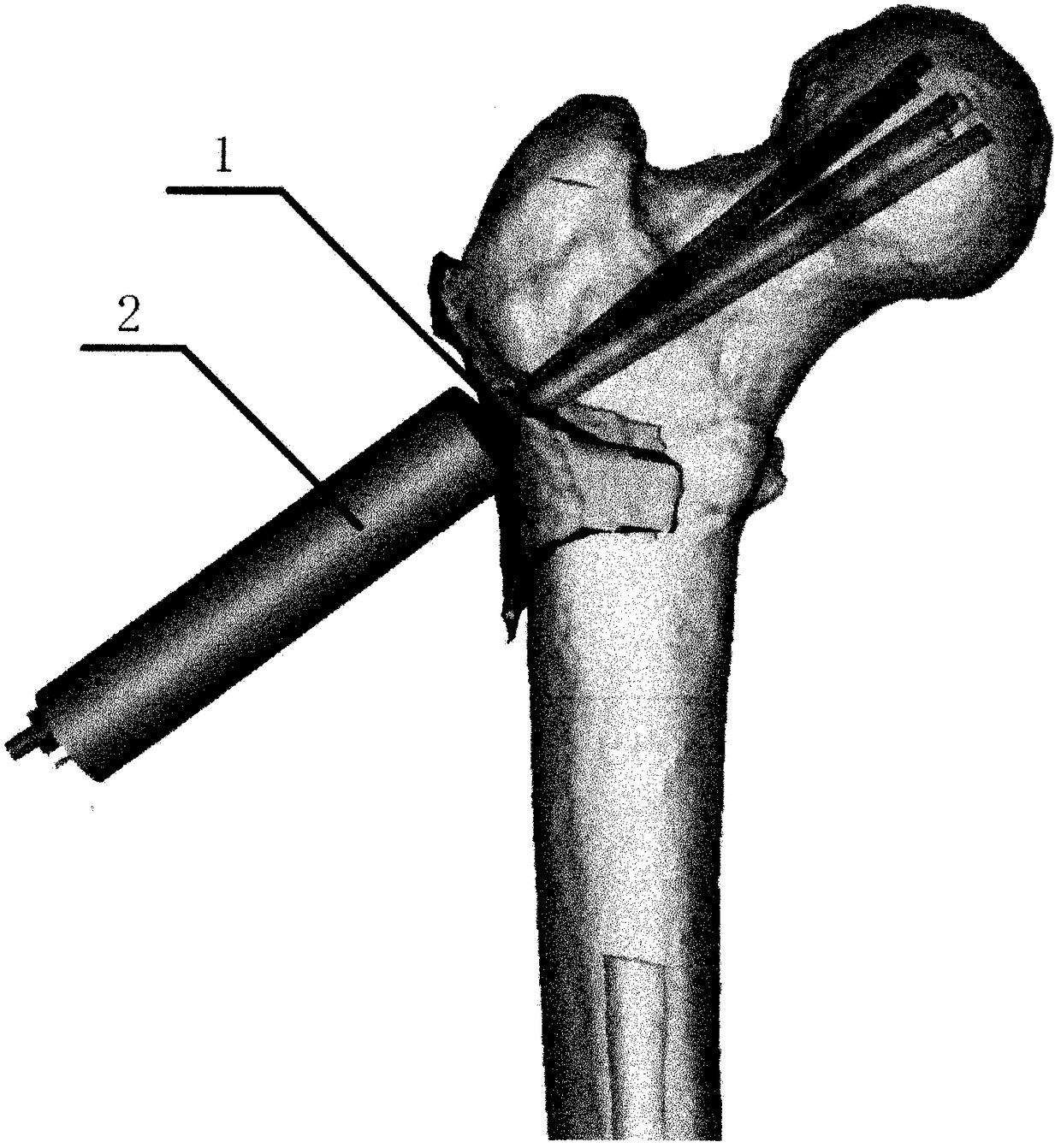
[0047] After determining the anatomical parameters of the normal proximal femur, according to the limitations of the parameters, the CDA software was used to first set up three digital samples of the guide with different arrangements of the decompression holes, and then the guide model was made by a 3D printer and tested on the normal femur model. accuracy. After confirming that the guide is working properly, the medical stainless steel metal guide and matching drill can be designed according...


PUM


Abstract

The invention provides a medullary core three-dimensional decompressing device for treating avascular necrosis of femoral head as well as preparing and testing methods based on the medullary core three-dimensional decompressing device, relating to the technical field of decompressing of the moral head. The medullary core three-dimensional decompressing device for treating avascular necrosis of the femoral head comprises a guide template, wherein the guide template is saddle-shaped in the whole, the inner side curve of the guide template attaches to the bone surface below the greater trochanter at the proximal femoral, a cylinder is arranged at the outer side of the guide template, a plurality of guide channels are arranged in the cylinder in a penetrating manner, and a plurality of through holes communicating with the guide channels are correspondingly formed in the guide template. The medullary core three-dimensional decompressing device can rapidly complete the multi-channel three-dimensional decompression for the necrotic femoral head.

Description

technical field [0001] The invention relates to the technical field of decompression of the femoral head, in particular to a preparation and testing method of a three-dimensional decompression device for ischemic necrosis of the femoral head. Background technique [0002] Osteonecrosis of the femoral head (ONFH), also known as avascular necrosis of the femoral head (AVNFH), is a common orthopaedic disease, and the definition of osteonecrosis of the femoral head (ONFH) by the International Society of Osteocirculation (ARCO) and the American Academy of Physicians (AAOS): ONFH is a disease in which the blood supply of the femoral head is interrupted or damaged, causing the death of bone cells and bone marrow components and subsequent repair, which in turn leads to structural changes of the femoral head, collapse of the femoral head, joint pain and joint dysfunction in patients. It is a common disease in the field of orthopedics. intractable disease. ARCO staging (M.Schmitt-Sod...

Claims


Application Information

Patent Timeline
Patent Type & Authority Patents(China)
IPC IPC(8): A61B17/17A61B34/10
CPCA61B17/17A61B17/1739A61B2017/00712A61B2017/00716
Inventor 王治
Owner THE AFFILIATED HOSPITAL OF SOUTHWEST MEDICAL UNIV
Why Eureka
Try Eureka