Novel in-vitro and in-vivo infection model based on Ebola virus-like particles
An Ebola virus, virus-like technology, applied in the biological field, can solve the problems of hindering the formation of virus-like particles, high price, and low throughput of β-lactamase activity measurement
- Summary
- Abstract
- Description
- Claims
- Application Information
AI Technical Summary
Problems solved by technology
Method used
Image
Examples
preparation example Construction
[0164] Preparation and infection experiment of EBOVLP
[0165] To prepare EBOVLP / Fluc, pcDNA-EBOV GP, pcDNA-EBOV VP40, pcDNA-EBOV NP and pcDNA-Fluc were co-transfected into 293T cells (purchased from the Cell Bank of Chinese Academy of Sciences) using PEI, cultured at 37°C for 4 hours, and the medium was changed to It was complete DMEM containing 10 μM sodium butyrate. After continuing to culture for 8 hours, the medium was replaced with FS293. After 48 hours, the supernatant was harvested and centrifuged at high speed to remove cell debris. The same method was used to prepare EBOVLP / Fluc / GP-, that is, the VLP without GP protein was used as a negative control.
[0166] 24 hours before the EBOVLP infection experiment, in a 96-well plate, add 1×10 4 Vero or Huh7.5.1 cells. After quantifying the VP40 contained in the supernatant VLP by Western blot, add 150ng of VP40-containing VLP to each well, and after incubation for a certain period of time, discard the supernatant, wash th...
Embodiment 1
[0185] Example 1 EBOVLP packaging of reporter gene protein
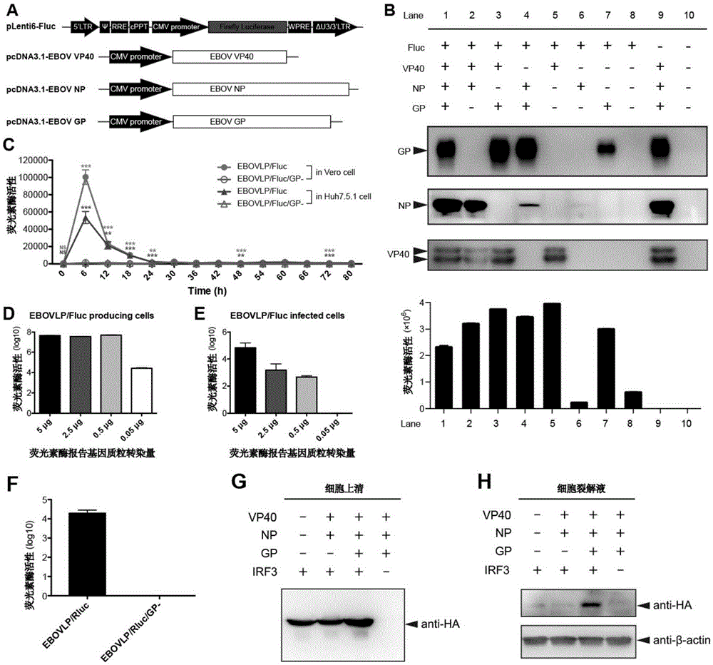
[0186] In order to express EBOVLP containing Fluc (EBOVLP / Fluc), the inventors constructed plasmids encoding Fluc, VP40, NP and GP ( figure 1 A), and these plasmids were co-transfected into 293T cells in different combinations, and the expression of VLP in the supernatant was detected by Western blot. Such as figure 1 As shown in B, co-transfected cells (lane 1) with plasmids encoding VP40, NP, GP and Fluc, all three viral proteins were expressed in the cells and released into the cell supernatant, and Fluc was detected in the supernatant However, when Fluc was expressed alone, Fluc could not be secreted into the supernatant (lane 8), which indicated that Fluc protein was successfully packaged into VLP and released out of the cell along with VLP. Since VP40 can be assembled into virus-like particles alone, so long as VP40 and Fluc (lane 5) are expressed at the same time, no matter whether it is co-expressed with NP...
Embodiment 2
[0187] Example 2 EBOVLP introduces the encapsulated reporter gene protein into cells through infection
[0188] Although VP40 alone could also encapsulate Flu in VLPs, when the inventors infected cells with supernatants containing intact granular EBOVLP / Fluc (lane 1) and granular EBOVLP / Fluc / GP-(lane 2) lacking the GP protein When , EBOVLP / Fluc can infect cells and release Fluc into the cells, resulting in the intracellular Fluc signal peaking at 6h after infection, while EBOVLP / Fluc / GP- is not infectious due to the lack of GP protein ( figure 1 C). Although the Fluc released into the cells was rapidly degraded after 6h, obvious Fluc signals could still be detected in EBOVLP / Fluc-infected cells at 48h and 72h after infection, which was significantly different from that in EBOVLP / Fluc / GP-infected cells. difference.
[0189] Then the inventors detected the activity of Fluc in 293T cells after transfection, hoping to determine the relationship between the amount of Fluc express...
PUM
| Property | Measurement | Unit |
|---|---|---|
| diameter | aaaaa | aaaaa |
Abstract
Description
Claims
Application Information
 - R&D
- Intellectual Property
- Life Sciences
- Materials
- Tech Scout
- Unparalleled Data Quality
- Higher Quality Content
- 60% Fewer Hallucinations
Browse by: Latest US Patents, China's latest patents, Technical Efficacy Thesaurus, Application Domain, Technology Topic, Popular Technical Reports.
© 2025 PatSnap. All rights reserved.Legal|Privacy policy|Modern Slavery Act Transparency Statement|Sitemap|About US| Contact US: help@patsnap.com


