Platelet-targeted microfluidic separation of cells

A technology for fluid separation and platelets, applied in microcarriers, animal cells, fluid controllers, etc.

Inactive Publication Date: 2020-12-29
THE GENERAL HOSPITAL CORP
View PDF25 Cites 0 Cited by
  • Summary
  • Abstract
  • Description
  • Claims
  • Application Information

AI Technical Summary

Problems solved by technology

Moreover, tumor cells have been reported to have active interactions with host cells in the microenvironment (e.g., blood cells such as leukocytes and platelets) during metastasis development, which may make the detection and isolation of CTCs more challenging

Method used


Image

  • Platelet-targeted microfluidic separation of cells
  • Platelet-targeted microfluidic separation of cells
  • Platelet-targeted microfluidic separation of cells

Examples


Embodiment 1

[0099] Example 1—Isolation of CTCs from Clinical Samples

[0100] This method was tested to determine its efficiency in isolating CTCs from different types of cancer.

[0101] Patients with advanced lung cancer, breast cancer, and melanoma were recruited according to an institutional review board (TRB)-approved protocol. All specimens were collected in the anticoagulant EDTA (Becton-Dickinson) tubes and process them using a microfluidic chip within 4 hours of blood draw. Additional platelet inhibitors such as theophylline, adenosine, dipyridamole, argatroban, and prostaglandin I2 were added immediately after blood collection. Collect fixed blood samples directly into BCT tube (Streck).

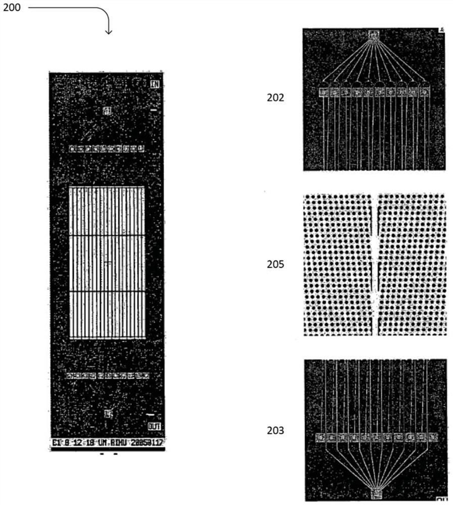
[0102] Samples were run on the two-stage microfluidic system described above. In particular, first-stage hydrodynamic sorting chips were fabricated on silicon wafers using deep reactive ion etching. Seal the chip using an anodically bonded glass lid to form a microfluidic chamber. A...

Embodiment 2

[0107] Example 2 - Isolation of different types of cancer

[0108] The microfluidic platform was extended to isolate CTCs from different types of cancer patients with epithelial (breast) and non-epithelial (melanoma) tumor origin. Breast CTCs were identified using the same protocol as for samples from lung cancer patients, but melanoma CTCs were stained with a cocktail of antibodies targeting melanoma-specific antigens (CSPG4, MCAM, TYRP1, and α-SMA) as previously reported. Preliminary results demonstrated reliable CTC capture for both cancer types (3 out of 5 breast samples with counts ranging from 0.9 to 2.7 CTC / mL; 5 of 6 melanoma samples with counts ranging from 1.4 to 17CTC / mL). Similar to the results in lung cancer, all CTCs captured using the current method were associated with varying degrees of platelet coverage.

[0109] As discussed above, lung and breast CTCs were identified using EpCAM / cadherin-11 (green) as epithelial and mesenchymal markers, but a cocktail o...

Embodiment 3

[0111] Example 3 - Anticoagulant and CTC Purity

[0112] A key issue potentially limiting the performance of platelet-targeting approaches is the relatively low purity of CTCs compared to other techniques. A large number of contaminating WBCs (>10 5 / mL), which makes downstream analysis very challenging. Staining with a platelet-specific antibody showed that most of the captured WBCs were also coated with platelets. The formation of these so-called platelet-leukocyte aggregates (PLAs) results from spontaneous platelet activation and subsequent expression of p-selectin, which will then bind to the PSGL-1 receptor on the surface of leukocytes. PLA cannot be removed by size-based sorting, and it is captured by anti-platelet antibodies along with platelet-associated CTCs.

[0113] To reduce the contamination of WBCs and improve the purity of CTCs, we tested a variety of platelet inhibitors for blood stabilization, such as theophylline, adenosine, dipyridamole, argatroban, and...


PUM

PropertyMeasurementUnit
sizeaaaaaaaaaa

Abstract

The present application relates to methods for isolating platelet-associated nucleated target cells, such as circulating epithelial cells, circulating tumor cells (CTCs), circulating endothelial cells (CECs), from sample fluids, such as biological fluids, such as blood, bone marrow, pleural effusion and ascitic fluid. ), circulating stem cells (CSCs), neutrophils and macrophages. The method comprises obtaining a cell capture chamber comprising a plurality of binding moieties bound to one or more walls of the chamber, wherein the binding moieties specifically bind platelets; upon binding the binding moieties to any platelet-associated nucleated target cells in the sample flowing the sample fluid through the cell capture chamber under binding conditions to form a complex; and separating and collecting platelet-associated nucleated target cells from the complex.

Description

[0001] Cross References to Related Applications [0002] This application claims priority to US Provisional Application Serial No. 62 / 034,522, filed August 7, 2014, the entire contents of which are incorporated herein by reference. [0003] Statement Regarding Federally Funded Research [0004] This invention was made with government support under Numbers 5-U01-EB012493-04 and EB002503-06A1 awarded by the National Institutes of Health, and 125929-PF-14-137-01-CCE awarded by the American Cancer Society . The government has certain rights in this invention. technical field [0005] The present invention relates to the isolation of nucleated cells from fluids such as blood. Background technique [0006] Metastasis, spread and growth of tumor cells from the primary site to distant organs represents the most devastating and lethal attribute of cancer and is responsible for 90% of cancer deaths. Although a systematic understanding of metastasis biology has not yet been establi...

Claims


Application Information

Patent Timeline
Patent Type & Authority Patents(China)
IPC IPC(8): G01N1/18G01N1/40G01N33/48G01N33/49G01N33/543G01N33/569G01N33/574
CPCC12N5/0694C12N2531/00B01L3/502761B01L2200/0652B01L2300/0864B01L2300/087B01L2300/0877B01L2400/086G01N33/54366G01N33/57488
Inventor M.托纳S.斯托特E.雷亚泰吉姜晓成
Owner THE GENERAL HOSPITAL CORP
Why Eureka
Try Eureka