Medical record image modeling system based on medical image

An image modeling and medical imaging technology, applied in medical image data management, medical informatics, medical data mining, etc.

Active Publication Date: 2017-06-27
CHENGDU GOLDISC UESTC MULTIMEDIA TECH
View PDF8 Cites 4 Cited by
  • Summary
  • Abstract
  • Description
  • Claims
  • Application Information

AI Technical Summary

Problems solved by technology

At the same time, the existing technology does not perform special treatment on the lesion/target ar...

Method used


Image

  • Medical record image modeling system based on medical image

Examples


Embodiment 1

[0059] Embodiment 1 is the splitting of the lobe of the brain; in this embodiment, the organ to be split is the lobe of the brain, and the multi-regions are the frontal lobe, the temporal lobe, the parietal lobe, the occipital lobe and the cerebellum;

[0060] The thin-layer scan image acquisition module is used to acquire a thin-layer scan image of T1-weighted imaging of the brain lobe;

[0061] T1-weighted imaging (T1-weighted imaging, T1WI) refers to this imaging method that focuses on the difference in longitudinal relaxation of tissues, while minimizing the influence of other tissue characteristics such as transverse relaxation on the image.

[0062] Described three-dimensional modeling module comprises:

[0063] Preprocessing unit: used to perform scalp and bone removal on the thin-layer scan image of the brain lobe;

[0064] Head model construction unit: used to construct a head model based on the preprocessed thin-slice scan image; the construction of the head model i...

Embodiment 2

[0071] Embodiment 2 is the splitting of the liver; in this embodiment, the organ to be split is the liver, and the multiple regions are the left lobe and the right lobe of the liver;

[0072] The thin-layer scan image acquisition module is used to read the DICOM sequence images of the liver using DCMTK;

[0073] Since the image storage and transmission of current medical imaging equipment is gradually moving closer to the DICOM standard, in the process of medical image processing, we often need to write various program modules related to DICOM format images to complete our own processing functions. It is a huge project to understand the DICOM protocol from scratch, and then write these codes to implement these protocols completely. The DCMTK developed by German offis company provides us with a platform to implement the DICOM protocol, so that we can easily complete our main work on the basis of it, without having to put too much energy on the details of the implementation of t...

Embodiment 4

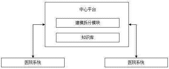
[0090] Embodiment 4 is that the hospital has its own internal database and data processing center, specifically: the data center is set in the hospital and is connected to multiple doctor terminals in the hospital through the intranet. Each doctor's terminal connected to the thin-layer scanning instrument is connected to the data center inside the hospital through the intranet; the data center inside the hospital processes and saves the data inside the hospital. When the doctor needs a model, he can directly download send. Intranet connection is used to improve security performance.

[0091] Further, in the above-mentioned embodiment, the doctor terminal can be a PC or a mobile terminal, both of which need to be configured with a corresponding client (C / S) or serve through a browser (B / S); virtual reality operation The device is a virtual reality glasses kit.

[0092]In addition, in the above-mentioned embodiment, the multi-region split model is operated through the virtual ...


PUM


Abstract

The invention discloses a medical record image modeling system based on a medical image. The system includes a central platform and a plurality of hospital systems connected with the central platform; the central platform includes a modeling and division module and a knowledge base, the central platform obtains thin-layer scan images, sent from the hospital systems, of organs to be modeled and divided, the modeling and division module conducts modeling and organ region division on the thin-layer scan images of the organs, and organ models obtained after modeling and division are stored in the form of disease cases in the knowledge base and then issued to the corresponding hospital systems. The hospital systems upload the thin-layer scan images to the central platform, the central platform conducts modeling and division on the images and stores the images, and therefore the medical record image modeling system is convenient for different hospitals or different doctors to obtain data.

Description

technical field [0001] The invention relates to a medical image-based medical record image modeling system. Background technique [0002] Medical imaging refers to the technology and processing process of obtaining internal tissue images of the human body or a certain part of the human body in a non-invasive manner for medical treatment or medical research. It includes the following two relatively independent research directions: medical imaging system and medical image processing. The former refers to the process of image formation, including the research on imaging mechanism, imaging equipment, imaging system analysis and other issues; the latter refers to the further processing of the obtained image, the purpose of which is to restore the original image that is not clear enough , or to highlight some feature information in the image, or to classify the pattern of the image and so on. [0003] The formation methods of medical images in the prior art include CT (Computed ...

Claims


Application Information

Patent Timeline
IPC IPC(8): G06F19/00
CPCG06F19/321G16H50/70
Inventor 曲建明曲飞寰范计朋
Owner CHENGDU GOLDISC UESTC MULTIMEDIA TECH
Why Eureka
Try Eureka